and Vincent L. Sorrell2
(1)
Division of Nuclear Medicine and Molecular Imaging Department of Radiology, University of Kentucky, Lexington, Kentucky, USA
(2)
Division of Cardiovascular Medicine Department of Internal Medicine Gill Heart Institute, University of Kentucky, Lexington, Kentucky, USA
Electronic supplementary material
The online version of this chapter (doi: 10.1007/978-3-319-25436-4_20) contains supplementary material, which is available to authorized users.
Given that hepatobiliary clearance is the predominant pathway for the 99mTc MPI radiopharmaceuticals, it is expected that the components of the biliary system (intrahepatic ducts, common hepatic duct, common bile duct) might be visualized and that the gallbladder will be visualized (Figs. 20.1 and 20.2) (Shih et al. 2002). A normal common bile duct is not uncommonly visible and should not be misconstrued as pathologic. While there are relatively few biliary conditions (biliary ectasia, common bile duct obstruction) that can be seen on SPECT MPI, there are multiple physiologic and pathologic conditions of the gallbladder (Table 20.1) (Hesse et al. 2005).


Fig. 20.1
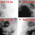
Normal intrahepatic bile ducts and common bile duct. The non-dilated bile ducts are prominent (a, b). Note absence of a gallbladder (cholecystectomy) (b). The left kidney is atrophic (a, c) in this patient on dialysis for end-stage kidney disease.(a) Stress raw projection images (Video 20.1a, frame 1), 99mTc sestamibi. (b) Stress raw projection image (Video 20.1b, frame 6), 99mTc sestamibi, right hepatic duct (one green line), left hepatic duct (two green lines), common bile duct (three green lines), gallbladder fossa (green circle). (c) Stress raw projection images (Video 20.1b, frame 47), 99mTc sestamibi, left kidney (yellow box)
Fig. 20.2
Contracted gallbladder in patient with dilated cardiomyopathy. The gallbladder is small and just seen “at the edge of the field-of-view” (a, b). Note the confluence of biliary ducts at the porta hepatis (a, c). The heart is enlarged with a dilated left ventricular cavity (a).(a) Stress raw projection images (Video 20.2a, frame 1), 99mTc sestamibi. (b) Stress raw projection image (Video 20.2b, frame 2), 99mTc sestamibi, gallbladder (green circle). (c) Stress raw projection image (Video 20.2b, frame 7), 99mTc sestamibi, porta hepatis bile ducts (green box)
Table 20.1
Differential diagnosis of “hot” and “cold” imaging findings related to the biliary system and gallbladder
Organ system | “Hot” finding | “Cold” finding | References |
|---|---|---|---|
Biliary system and gallbladder | Biliary ectasia with stasis Common bile duct obstruction | Inadequate or prolonged fasting Cholecystitis (acute , chronic) Biliary stricture/stone at ampulla of Vater | Chamarthy and Travin (2010) Gedik et al. (2007) Hesse et al. (2005) Howarth et al. (1996) Meesala et al. (2006) Panjrath et al. (2004) Toran et al. (1997) |
The gallbladder should fill with radioactive bile and appear quite “hot,” often the “hottest” finding in the field-of-view. The patient is typically fasting for the stress test, and the gallbladder, as in 99mTc iminodiacetic acid (IDA) hepatobiliary scintigraphy, should fill. Diaphragmatic motion may produce a moving (“beating”) gallbladder (Fig. 20.3). The gallbladder generally has a characteristic round to ovoid shape and is tucked under the inferior right lobe; however, it may be intrahepatic in location (Howarth et al. 1996). Figure 20.4 shows an unusually elongated and superiorly displaced gallbladder; it is adjacent to the right hemidiaphragm by anatomic CT correlation. A very “hot” and “high” gallbladder may create a processing artifact on the reconstructed MPI (Fig. 20.5). The degree of gallbladder visualization can vary from rest to stress imaging on both 1-day and 2-day protocols (Figs. 20.6 and 20.7). The gallbladder is usually well-distended and a contracted gallbladder is considered abnormal (Fig. 20.8).


Fig. 20.3
“Beating gallbladder.” Note how the gallbladder appears to be fluttering throughout both acquisitions (a, b); this is a clue to right hemidiaphragmatic breathing motion which could lead to motion artifact on processed SPECT MPI. The gallbladder should be stationary; however, the small intestinal activity should change with normal peristalsis as seen here (a, b).(a) Rest raw projection images (Video 20.3a, frame 1), 99mTc sestamibi. (b) Stress raw projection images (Video 20.3b, frame 1), 99mTc sestamibi
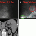
Fig. 20.4


Elongated, elevated gallbladder. Note the unusual shape and position of the gallbladder (a–c). The CT images define its anatomic position and morphology (d, e).(a) Rest raw projection images (Video 20.4a, frame 1), 99mTc sestamibi. (b) Stress raw projection images (Video 20.4b, frame 1), 99mTc sestamibi. (c) Stress raw projection image (Video 20.4c, frame 18), 99mTc sestamibi, gallbladder (green outline). (d) Coronal CT through more anterior liver and gallbladder, septate gallbladder abuts right hemidiaphragm (yellow box). (e) Coronal CT through more posterior liver and gallbladder, posteriormost tail of gallbladder (yellow box) abuts right hemidiaphragm
Related posts:

Stay updated, free articles. Join our Telegram channel
Full access? Get Clinical Tree
