and Vincent L. Sorrell2
(1)
Division of Nuclear Medicine and Molecular Imaging Department of Radiology, University of Kentucky, Lexington, Kentucky, USA
(2)
Division of Cardiovascular Medicine Department of Internal Medicine Gill Heart Institute, University of Kentucky, Lexington, Kentucky, USA
Electronic supplementary material
The online version of this chapter (doi: 10.1007/978-3-319-25436-4_28) contains supplementary material, which is available to authorized users.
28.1 Case Challenge #13
28.1.1 Problem
Clinical Highlights
A 59-year-old male presents for preoperative risk assessment.
Images for Review


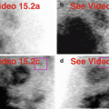
Fig. 28.1
(a) Rest raw projection images (Video 28.1a, frame 1),99mTc sestamibi
(b) Stress raw projection images (Video 28.1b, frame 1),99mTc sestamibi
(c) Stress/rest processed SPECT images (SA, HLA, VLA) (without and with AC)
(d) Stress and rest gated SPECT images (Video 28.1c, frame 1) (SA, VLA, HLA)
Characterize the Pertinent Finding(s)
Chest
Parathyroid glands: □ hot □ cold
Breasts: □ hot □ cold
Chest wall: □ hot □ cold
Skeleton: □ hot □ cold
Pleura: □ hot □ cold
Lungs: □ hot □ cold
Mediastinum: □ hot □ cold
Myocardium and pericardium: □ hot □ cold
Right atrium and right ventricle: □ hot □ cold
Abdomen
Peritoneum: □ hot □ cold
Liver: □ hot □ cold
Biliary system and gallbladder: □ hot □ cold
Spleen: □ hot □ cold
Stomach: □ hot □ cold
Small intestine and large intestine: □ hot □ cold
Adrenal glands: □ hot □ cold
Kidneys and female reproductive system: □ hot □ cold
State Your Relevant Diagnosis(es)
Chest
Parathyroid glands: □ normal □ mediastinal adenoma
Breasts: □ normal □ malignancy
Chest wall: □ normal □ arms by sides
Skeleton: □ normal □ anemia
Pleura: □ normal □ effusion
Lungs: □ normal □ emphysema
Mediastinum: □ normal □ malignancy
Myocardium and pericardium: □ normal □ effusion
Right atrium and right ventricle: □ normal □ pulmonary hypertension
Abdomen
Peritoneum: □ normal □ ascites
Liver: □ normal □ cirrhosis
Biliary system and gallbladder: □ normal □ prolonged fasting
Spleen: □ normal □ splenomegaly
Stomach: □ normal □ gastropathy
Small intestine and large intestine: □ normal □ large intestine across upper abdomen
Adrenal glands: □ normal □ malignancy
Kidneys and female reproductive system: □ normal □ polycystic disease
28.1.2 Solution
Additional Annotated Images
(e) Stress/rest processed SPECT images (SA, HLA, VLA) (without and with AC), right ventricle (yellow ovals) on selected SA and HLA images
The Pertinent Findings
Chest
Skeleton: ■ hot □ cold
Right atrium and right ventricle: ■ hot ■ cold
Abdomen
Peritoneum: □ hot ■ cold
Liver: □ hot ■ cold
Biliary system and gallbladder: ■ hot □ cold
Spleen: ■ hot □ cold
Stomach: ■ hot □ cold
The Relevant Diagnosis(es)
Chest
Skeleton: ■ anemia
Right atrium and right ventricle: ■ pulmonary hypertension
Abdomen
Peritoneum: ■ ascites
Liver: ■ cirrhosis
Biliary system and gallbladder: ■ normal
Spleen: ■ splenomegaly
Stomach: ■ gastropathy
Discussion
Chest
The right ventricle is enlarged and has a thickened “hot” wall (a, b, c, d, e). Note the flattened septum and D-shaped left ventricle. Function is preserved (d). The skeleton is slightly prominent, consistent with liver disease-related anemia.
Abdomen
The abdomen demonstrates the classical constellation of findings of advanced liver disease and portal hypertension: small liver, “cold” ascites, “hot” spleen, and “hot” stomach. Note the normally filled gallbladder. Most often, even in cirrhosis, the gallbladder will be visualized.
The intestinal tract and left kidney are not included in the field-of-view and cannot be evaluated.
Relevant Chapter(s)
8, 13, 18, 19, 20, 21, and 22
28.2 Case Challenge #14
28.2.1 Problem
Clinical Highlights
A 62-year-old female has chest pain.
Images for Review

Fig. 28.2
(a) Rest raw projection images (Video 28.2a, frame 1),99mTc sestamibi
(b) Stress raw projection images (Video 28.2b, frame 1),99mTc sestamibi
Characterize the Pertinent Finding(s)
Chest
Breasts: □ hot □ cold
Chest wall: □ hot □ cold
Skeleton: □ hot □ cold
Pleura: □ hot □ cold
Lungs: □ hot □ cold
Mediastinum: □ hot □ cold
Myocardium and pericardium: □ hot □ cold
Right atrium and right ventricle: □ hot □ cold
Vascular system: □ hot □ cold
Abdomen
Peritoneum: □ hot □ cold
Liver: □ hot □ cold
Biliary system and gallbladder: □ hot □ cold
Spleen: □ hot □ cold
Stomach: □ hot □ cold
Small intestine and large intestine: □ hot □ cold
Adrenal glands: □ hot □ cold
Kidneys and female reproductive system: □ hot □ cold
State Your Relevant Diagnosis(es)
Chest
Breasts: □ normal □ mastectomy
Chest wall: □ normal □ contamination artifact
Skeleton: □ normal □ anemia
Pleura: □ normal □ effusion
Lungs: □ normal □ pneumonectomy
Mediastinum: □ normal □ hilar mass
Myocardium and pericardium: □ normal □ myocardial mass
Right atrium and right ventricle: □ normal □ enlargement
Vascular system: □ normal □ port injection site
Abdomen
Peritoneum: □ normal □ ascites
Liver: □ normal □ cirrhosis
Biliary system and gallbladder: □ normal □ cholecystectomy
Spleen: □ normal □ splenectomy
Stomach: □ normal □ gastropathy
Small intestine and large intestine: □ normal □ ileus
Adrenal glands: □ normal □ mass
Kidneys and female reproductive system: □ normal □ renal cyst
28.2.2 Solution
Additional Annotated Images
None.
The Pertinent Findings
Chest
Not applicable
Abdomen
Peritoneum: □ hot ■ cold
Liver: □ hot ■ cold
Biliary system and gallbladder: □ hot ■ cold
Spleen: □ hot ■ cold
Stomach: ■ hot □ cold
The Relevant Diagnosis(es)
Chest
Not applicable
Abdomen
Peritoneum: ■ ascites
Liver: ■ cirrhosis
Biliary system and gallbladder: ■ cholecystectomy
Spleen: ■ splenectomy
Stomach: ■ gastropathy
Discussion
Chest
There are no abnormalities.
Abdomen
There is photopenia due to ascites throughout the abdomen (a, b). The liver is small, consistent with cirrhosis, and the stomach is diffusely “hot,” consistent with gastropathy. This constellation is characteristic of the sequelae of cirrhosis. The unusual feature to this case is absence of the spleen rather than the expected splenomegaly; according to the medical record, the spleen is surgically absent from unrelated trauma in the past. The gallbladder is also surgically absent. The left kidney is present.
Relevant Chapter(s)
18, 19, 20, 21, and 22
28.3 Case Challenge #15
28.3.1 Problem
Clinical Highlights
A 50-year-old male awaits renal transplant; he provides a previous surgical history.
Images for Review


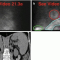
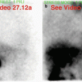
Fig. 28.3
(a) Current: stress raw projection images (Video 28.3a, frame 1),99mTc sestamibi (75 minutes after injection)
(b) Two years previously: stress raw projection images (Video 28.3b, frame 1),99mTc sestamibi
Characterize the Pertinent Finding(s)
Chest
Breasts: □ hot □ cold
Chest wall: □ hot □ cold
Skeleton: □ hot □ cold
Pleura: □ hot □ cold
Lungs: □ hot □ cold
Mediastinum: □ hot □ cold
Myocardium and pericardium: □ hot □ cold
Right atrium and right ventricle: □ hot □ cold
Vascular system: □ hot □ cold
Abdomen
Abdominal wall: □ hot □ cold
Peritoneum: □ hot □ cold
Liver: □ hot □ cold
Biliary system and gallbladder: □ hot □ cold
Spleen: □ hot □ cold
Stomach: □ hot □ cold
Small intestine and large intestine: □ hot □ cold
Adrenal glands: □ hot □ cold
State Your Relevant Diagnosis(es)
Chest
Breasts: □ normal □ gynecomastia
Chest wall: □ normal □ pacemaker
Skeleton: □ normal □ metastatic disease
Pleura: □ normal □ mesothelioma
Lungs: □ normal □ emphysema
Mediastinum: □ normal □ metastatic disease
Myocardium and pericardium: □ normal □ effusion
Right atrium and right ventricle: □ normal □ pulmonary hypertension
Vascular system: □ normal □ injection site
Abdomen
Abdominal wall: □ normal □ patient’s arm and hand
Peritoneum: □ normal □ ascites
Liver: □ normal □ transplant for cirrhosis
Biliary system and gallbladder: □ normal □ cholecystectomy
Spleen: □ normal □ enlargement
Stomach: □ normal □ gastric outlet obstruction
Small intestine and large intestine: □ normal □ large bowel visualization
Adrenal glands: □ normal □ metastatic lung cancer
28.3.2 Solution
Additional Images
(c) Current: stress raw projection images, contrast adjusted for breasts (Video 28.3c, frame 1),99mTc sestamibi (75 minutes after injection)
The Pertinent Findings
Chest
Breasts: ■ hot □ cold
Abdomen
Liver: ■ hot □ cold
Biliary system and gallbladder: □ hot ■ cold
Spleen: ■ hot □ cold
Small intestine and large intestine: ■ hot □ cold
The Relevant Diagnosis(es)
Chest
Breasts: ■ gynecomastia
Abdomen
Liver: ■ transplant for cirrhosis
Biliary system and gallbladder: ■ cholecystectomy
Spleen: ■ enlargement
Stomach: ■ normal
Small intestine and large intestine: ■ large bowel visualization
Discussion
Chest
There is subtle, symmetric uptake in the areolar breast tissue in this male patient who underwent liver transplant for cirrhosis; this suggests gynecomastia (a, b, c).
Abdomen
The liver appears normal on the current examination (a), but it was distinctly abnormal on the previous examination before the transplant (b). Note difference in liver size, shape, and uptake pattern – and he no longer has a gallbladder! The enlarged spleen (a, b) persists after transplant (a, b). Prominent large intestinal activity on the current examination (a) could be mistaken for a normal gallbladder; follow the activity on the cinematic display.
Relevant Chapter(s)
6, 19, 20, 21, and 23
28.4 Case Challenge #16
28.4.1 Problem
Clinical Highlights
A 58-year-old diabetic female with known coronary artery disease undergoes stress testing to exclude inducible ischemia. She has chronic obstructive pulmonary disease and provides a past surgical history following a motor vehicle accident.
Images for Review


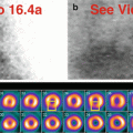
Fig. 28.4
(a) Rest raw projection images (Video 28.4a, frame 1),99mTc sestamibi
(b) Stress raw projection images (Video 28.4b, frame 1),99mTc sestamibi
(c) Stress/rest processed SPECT images (SA, HLA, VLA) (with and without AC)
Characterize the Pertinent Finding(s)
Chest
Thyroid gland: □ hot □ cold
Breasts: □ hot □ cold
Chest wall: □ hot □ cold
Pleura: □ hot □ cold
Lungs: □ hot □ cold
Mediastinum: □ hot □ cold
Myocardium and pericardium: □ hot □ cold
Right atrium and right ventricle: □ hot □ cold
Diaphragm: □ hot □ cold
Abdomen
Abdominal wall: □ hot □ cold
Liver: □ hot □ cold
Biliary system and gallbladder: □ hot □ cold
Spleen: □ hot □ cold
Stomach: □ hot □ cold
Small intestine and large intestine: □ hot □ cold
Kidneys and female reproductive system: □ hot □ cold
Vascular system: □ hot □ cold
State Your Relevant Diagnosis(es)
Chest
Thyroid gland: □ normal □ substernal goiter
Breasts: □ normal □ soft-tissue attenuation artifact
Chest wall: □ normal □ pacemaker
Pleura: □ normal □ effusion
Lungs: □ normal □ emphysema
Mediastinum: □ normal □ cystic mass
Myocardium and pericardium: □ normal □ effusion
Right atrium and right ventricle: □ normal □ right auricular appendage and pulmonary hypertension
Diaphragm: □ normal □ flattening
Abdomen
Abdominal wall: □ normal □ urinary contamination artifact
Liver: □ normal □ polycystic disease
Biliary system and gallbladder: □ normal □ dilated intrahepatic ducts with stasis
Spleen: □ hot □ cold
Stomach: □ normal □ fluid filled after ingestion of fluids
Small intestine and large intestine: □ normal □ internal hernia
Kidneys and female reproductive system: □ normal □ hydronephrotic obstructed left kidney
Vascular system: □ normal □ extravasation
28.4.2 Solution
Additional Annotated Images
(d) PA chest radiograph
(e) Stress raw projection image (Video 28.4c, frame 9, Anterior, image 9),99mTc sestamibi, right auricular appendage (blue oval)
(f) Stress raw projection image (Video 28.4c, frame 60, left lateral, image 60),99mTc sestamibi, “cold” stomach with “hot” wall (orange lines)
The Pertinent Findings
Chest
Breasts: □ hot ■ cold
Lungs: □ hot ■ cold
Right atrium and right ventricle: ■ hot ■ cold
Diaphragm: □ hot ■ cold
Abdomen
Spleen: □ hot ■ cold
Stomach: ■ hot ■ cold
The Relevant Diagnosis(es)
Chest
Breasts: ■ soft-tissue attenuation artifact
Lungs: ■ emphysema
Right atrium and right ventricle: ■ right auricular appendage and pulmonary hypertension
Diaphragm: ■ flattening
Abdomen
Spleen: ■ splenectomy
Stomach: ■ fluid filled after ingestion of fluids
Discussion
Chest
There is superimposed “cold” breast tissue (a, b) which creates slight breast attenuation artifact in the anterior wall of the left ventricle (c). There are hyperexpanded “cold” lungs, especially involving the lower lobes, with flattened diaphragms (a, b); radiography is confirmatory (d).
The right ventricle is enlarged with prominent right ventricular myocardial uptake (a, b, e), consistent with pulmonary hypertension in the setting of advanced obstructive pulmonary disease; there is a normal right auricular appendage (a, b, e). There is a large reversible perfusion defect in the inferior wall consistent with right coronary artery ischemia (c).
Abdomen
The biliary system appears normal; a gallbladder is present but is not included in the field-of-view. The spleen is surgically absent, and the left kidney and small intestine occupy the left upper quadrant adjacent to the stomach (a, b). The stomach appears both “hot” and “cold,” likely reflecting gastric wall uptake, plus a mixture of duodenogastric reflux and fluid ingestion (a, b, f). Given the history of diabetes mellitus, prolonged gastric distension can suggest gastroparesis.
Relevant Chapter(s)
6, 10, 13, 16, 21, and 22
28.5 Case Challenge #17
28.5.1 Problem
Clinical Highlights
A 33-year-old male undergoes preoperative planning. He complains of dyspnea. The regadenoson stress test is normal. Recent echocardiogram showed LVEF of 67 %. Calcified gallstones are noted on ultrasonography.
Related posts:

Stay updated, free articles. Join our Telegram channel

Full access? Get Clinical Tree


