and Vincent L. Sorrell2
(1)
Division of Nuclear Medicine and Molecular Imaging Department of Radiology, University of Kentucky, Lexington, Kentucky, USA
(2)
Division of Cardiovascular Medicine Department of Internal Medicine Gill Heart Institute, University of Kentucky, Lexington, Kentucky, USA
Electronic supplementary material
The online version of this chapter (doi: 10.1007/978-3-319-25436-4_22) contains supplementary material, which is available to authorized users.
Table22.1 lists various causes for a “hot” or “cold” stomach on MPI. Gastric activity can be quite confounding with respect to reconstruction processing artifacts affecting the inferior myocardial wall. Most often, gastric activity is related to intraluminal duodenogastric bile reflux that may occur to different degrees of severity between patients and between rest and stress imaging in the same patient (Fig.22.1). Gastric activity obscures the inferior myocardial wall and frequently creates processing artifacts. Ideally, subdiaphragmatic gastric activity is avoided altogether (Burrell and MacDonald2006; Middleton and Williams1994).
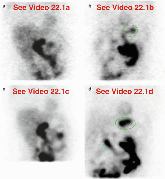
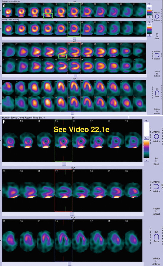
Table 22.1
Differential diagnosis of “hot” and “cold” imaging findings related to the stomach
Organ system | “Hot” finding | “Cold” finding | References |
|---|---|---|---|
Stomach | Duodenogastric reflux Gastroparesis Gastropathy (dyspepsia/gastritis, cirrhosis) Free99mTc pertechnetate | Ingestion of fluids Distension, acute or chronic | Burrell and MacDonald (2006) Cote and Dumont (2004) Gedik et al. (2007) Gholamrezanezhad et al. (2006) Gupta et al. (2015) Khary et al. (1995) Middleton and Williams (1994) Middleton and Williams (1996) |
Fig. 22.1
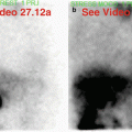
Variable degrees of intermittent duodenogastric reflux. On rest (a,b) and stress (c,d) imaging, there are different degrees (intensities) of gastric activity: on rest, there is much less with only a few episodes of duodenogastric reflux, while on stress, there is much more refluxed activity that persists longer. The variable appearance indicates intraluminal activity; gastric wall uptake will not change between rest and stress. Note that gastric activity is clearly apparent only on stress processed SPECT and gated SPECT images (e,f); there is no significant artifactual effect on the adjacent inferior myocardial wall. A previous cholecystectomy is the reason for non-visualization of the gallbladder (a,c).(a) Rest raw projection images (Video 22.1a, frame 1),99mTc sestamibi. (b) Rest raw projection image (Video 22.1b, frame 31),99mTc sestamibi, gastric activity (green oval). (c) Stress raw projection images (Video 22.1c, frame 1),99mTc sestamibi. (d) Stress raw projection image (Video 22.1d, frame 24),99mTc sestamibi, gastric activity (green oval), duodenal activity (blue lines). (e) Stress/rest processed SPECT images (SA, VLA, HLA), gastric activity (green box) on selected SA and VLA images. (f) Stress gated SPECT images (Video 22.1e, frame 1) (SA, VLA, HLA)
Drinking, eating, changing to a prone position, waiting, and repeating MPI are often the most practical maneuvers to lessen this common artifact (Fig.22.2) (Burrell and MacDonald2006). Refluxed duodenogastric activity may persist within the gastric lumen for a long time in patients with gastroparesis; sometimes ingestion of water or food helps clear it (Fig.22.3), but at other times, there is no significant benefit (Gedik et al.2007). Patients with repeat examinations may show the same pattern (Fig.22.4); knowing this at the time of the follow-up MPI may prompt modifications to the imaging protocol such as a longer fasting state if possible, particularly in diabetics who are at risk to develop gastroparesis. In one series, three patients out of 76 had uninterpretable examinations (Middleton and Williams1996). Because the fundus is dependent, laying the patient with right side down for 20 minutes facilitated clearance of the stomach, resulting in all examinations being interpretable; the frequency and degree of duodenogastric reflux were considerably lower with the maneuver (Middleton and Williams1996). This maneuver might be an option for facilities with semi-upright/recumbent dedicated cardiac gamma camera systems that cannot be easily configured for prone image acquisition.
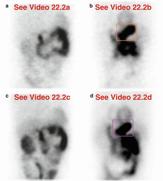
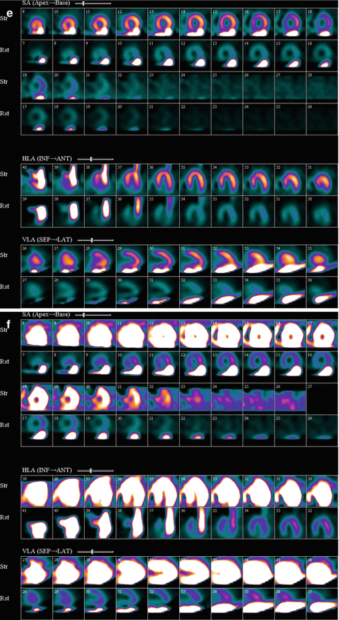
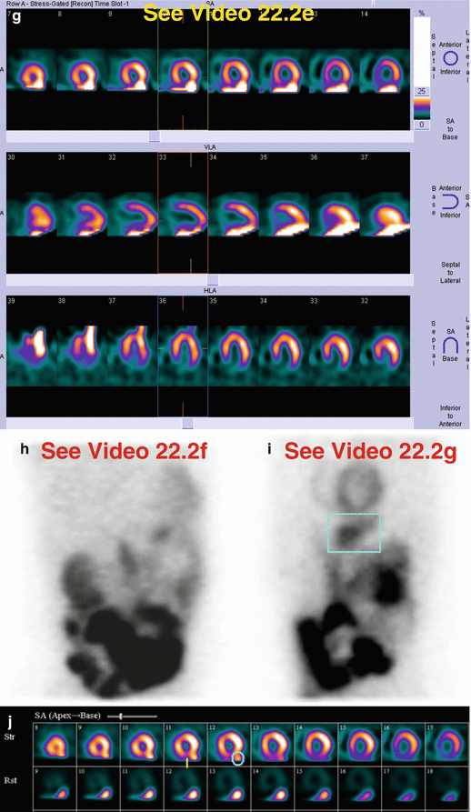
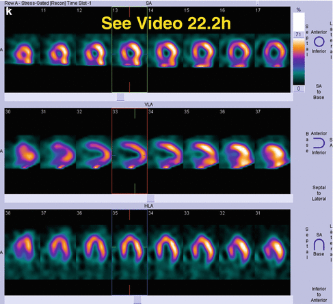
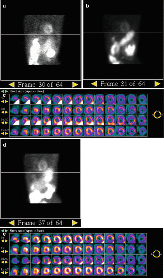
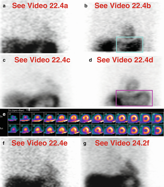
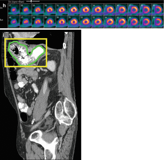
Fig. 22.2
Problematic gastric activity, clearing after water and waiting. This 60-year-old male has a history of previous myocardial infarction and hypertension; he underwent a regadenoson stress test. The “hot” stomach on the initial rest (a,b) and stress (c,d) images creates a significant defect in the inferior myocardium (e–g). The gastric activity clears on the repeat stress images (h,i) after water and waiting, and there is improvement in the artifact (j,k). There is an apparent small, mild perfusion defect in the inferior wall, but its characterization is compromised by the adjacent gastric activity; it was interpreted as a possible scar or processing artifact. On the gated SPECT images (g,k), the inferior wall shows mild hypokinesis, but its evaluation is limited by the gastric activity. Visually, left ventricular function was low normal.(a) Rest raw projection images (Video 22.2a, frame 1),99mTc sestamibi. (b) Rest raw projection image (Video 22.2b, frame 36),99mTc sestamibi, gastric activity (orange box). (c) Initial stress raw projection images (Video 22.2c, frame 1),99mTc sestamibi. (d) Initial stress raw projection image (Video 22.2d, frame 38),99mTc sestamibi, gastric activity (pink box). (e) Initial stress/rest processed SPECT images optimized for stress images (SA, HLA, VLA). (f) Initial stress/rest processed SPECT images optimized for rest images (SA, HLA, VLA). (g) Initial stress gated SPECT images (Video 22.2e, frame 1) (SA, VLA, HLA). (h) Repeat stress raw projection images after water and 20 minute wait (Video 22.2f, frame 1),99mTc sestamibi. (i) Repeat stress raw projection image after water and 20 minute wait (Video 22.2g, frame 39),99mTc sestamibi, gastric activity (blue box). (j) Repeat stress/rest processed SPECT images optimized for stress images (SA), gastric activity (blue oval), inferior wall defect (vertical yellow line). (k) Repeat stress gated SPECT images (Video 22.2h, frame 1) (SA, VLA, HLA)
Fig. 22.3
Diabetic gastroparesis, minimal clearing of problematic gastric activity with water. There is a “hot” stomach on the initial stress images (a–c). It is partially mitigated by re-imaging after water (d,e); the stomach may empty slowly in functional motility disorders. There is a small inferior wall defect on rest images; the inferior and inferolateral walls from apex to base are almost impossible to evaluate on initial stress images (c), and only somewhat evaluable on repeat stress images (e).(a) Selected rest raw projection image,99mTc tetrofosmin, level of inferior wall (white line). (b) Selected stress raw projection image,99mTc tetrofosmin, level of inferior wall (white line). (c) Stress/rest processed SPECT images (SA) (without and with AC). (d) Selected repeat stress raw projection image 90 minutes after water ingestion, level of inferior wall (white line). (e) Repeat stress/original rest processed SPECT images (SA) (without and with AC)
Fig. 22.4


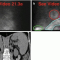
Nondiabetic gastroparesis with “hot” and “cold” stomach. The current MPI demonstrates a distended “cold” stomach with a “hot” wall on both rest (a,b) and stress (c,d) raw projection images. Although the gastric activity is clearly apparent on the processed images (e), there is only a small, mild, fixed inferoapical defect. Interestingly, the raw data patterns are similar to those seen four years earlier (f,g); at that time, the processed data did not demonstrate significant artifact from the gastric activity (h). Although stomach-related artifactual effects are not always predictable, as shown here, comparison with previous images and reports can guide follow-up imaging techniques. Note the stomach morphology; it extends from anterior to posterior and lies immediately inferior to the heart (i).(a) Rest raw projection images (Video 22.4a, frame 1),99mTc sestamibi. (b) Rest raw projection image (Video 22.4b, frame 31),99mTc sestamibi, stomach (blue box). (c) Stress raw projection images (Video 22.4c, frame 1),99mTc sestamibi. (d) Stress raw projection image (Video 22.4d, frame 38),99mTc sestamibi, stomach (pink box). (e) Stress/rest processed SPECT images (SA). (f) Four years earlier: rest raw projection images (Video 22.4e, frame 1),99mTc sestamibi. (g) Four years earlier: stress raw projection images (Video 22.4f, frame 1),99mTc sestamibi. (h) Four years earlier: stress/rest processed SPECT images (SA). (i) Sagittal CT image of the upper abdomen and stomach (outlined ingreen, withinyellow box)
Related posts:

Stay updated, free articles. Join our Telegram channel
Full access? Get Clinical Tree
