and Vincent L. Sorrell2
(1)
Division of Nuclear Medicine and Molecular Imaging Department of Radiology, University of Kentucky, Lexington, Kentucky, USA
(2)
Division of Cardiovascular Medicine Department of Internal Medicine Gill Heart Institute, University of Kentucky, Lexington, Kentucky, USA
Electronic supplementary material
The online version of this chapter (doi: 10.1007/978-3-319-25436-4_15) contains supplementary material, which is available to authorized users.
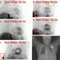
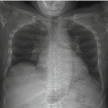
Table 15.1 lists benign and malignant conditions affecting the lymphatic system that may be seen during SPECT MPI. Axillary lymph nodes may be visualized, particularly if the arms are positioned “up.” As discussed in Chap. 14, extravasated injections result in lymphatic clearing with deposition in the axillary lymph nodes (Figs. 15.1 and 15.2) (Chamarthy and Travin 2010; Gedik et al. 2007; Gentili et al. 1994; Shih et al. 2002; Williams et al. 2003). At times, the lymphatic channels can be visualized as “hot” lines (Shih et al. 1999). Verifying extravasation is important so as not to casually attribute malignant uptake incorrectly to a benign reason.

Table 15.1
Differential diagnosis of “hot” and “cold” imaging findings related to the lymphatic system
Organ system | “Hot” finding | “Cold” finding | References |
|---|---|---|---|
Lymphatic system | Sarcoidosis Neoplasm, metastasis (e.g., breast carcinoma) Lymphoma Venous extravasation resulting in visualization of axillary lymph node(s) | Necrotic lymph node(s) Neoplasm, metastasis | Aktolun et al. (1994) Aktolun and Bayhan (1994) Chamarthy and Travin (2010) Gedik et al. (2007) Gentili et al. (1994) Mlikotic and Mishkin (2000) Shih et al. (2002) Shih et al. (1999) Taillefer et al. (1995) Taillefer et al. (1998) Waxman (1997) Williams et al. (2003) |
Fig. 15.1


Right axillary lymph node visualization. The right axilla appears normal on rest imaging (a). Extravasation at the right arm injection site during the stress test resulted in lymphatic uptake on stress imaging (b). Note how the right arm is raised on both acquisitions, permitting a clear view of the right axilla on the more right anterior oblique projections. Conversely, the left axilla is generally seen to better advantage on the more left anterior oblique projections (see Fig. 15.2).(a) Rest raw projection image, RAO, 99mTc tetrofosmin, inferior aspect of the heart (white line). (b) Stress raw projection image, RAO, 99mTc tetrofosmin, right axillary lymph node (yellow box)
Related posts:

Stay updated, free articles. Join our Telegram channel
Full access? Get Clinical Tree
