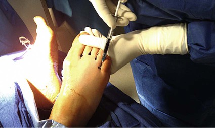
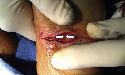
Linda Kelahan, Denis Primakov, Chad Baarson, Ali Noor and Anthony C. Venbrux Lymphangiography is rarely performed today because cross-sectional imaging to evaluate lymph node pathology has replaced it.1 Years ago, prior to ultrasound (US), computed tomography (CT), and magnetic resonance imaging (MRI), bipedal lymphangiography was used in part to stage malignancies (e.g., lymphomas and metastatic disease to lymph nodes).2,3 Injury to the thoracic duct (traumatic, surgical, etc.) may be associated with life-threatening chylous pleural effusions. Surgery to ligate the thoracic duct is associated with significant morbidity and mortality; such patients are frequently severely debilitated.4–7 Percutaneous thoracic duct embolization is an alternative technique and in some centers has changed the management of thoracic duct injuries. The key is opacification of the thoracic duct and percutaneous puncture of the duct in the abdomen. Once the thoracic duct has been accessed percutaneously, superselective catheterization of the duct requires use of a microcatheter and guidewire and experience in embolotherapy.8,9 The initial step is generally bipedal lymphangiography.1–3 The technique is briefly outlined here. In some centers, only the lymphatics of the right foot are accessed. Recent developments include ultrasound-guided direct percutaneous puncture of lymph node(s) in the femoral regions. This latter technique may save time, but experience using this approach is anecdotal and limited. In terms of equipment, a lymphangiography injector, once standard equipment in interventional radiology suites, is no longer manufactured. Use of a handheld insufflator (used for angioplasty) will substitute. The goal of this procedure is to opacify the cisterna chyli or other dominant lymphatics in the abdomen. Initially, 0.1 to 0.2 mL of 1% methylene blue is injected into each interspace between the toes of both feet using a 1-mL syringe (Fig. 123-1). Patients should be warned that their feet may remain blue for days to weeks, and their urine will also develop a blue-green coloration. After waiting approximately 10 minutes to allow the dye to be taken up by the lymphatics, the dorsum of each foot is then anesthetized with 1% lidocaine. A curved blade is then used to make a shallow transverse incision (≈2.5 cm) across the dorsum of each foot, taking care to only cut the epidermis and the topmost layer of the dermis (Fig. 123-2).
Bipedal Lymphangiography
Introduction
Technique
![]()
Stay updated, free articles. Join our Telegram channel

Full access? Get Clinical Tree


Radiology Key
Fastest Radiology Insight Engine