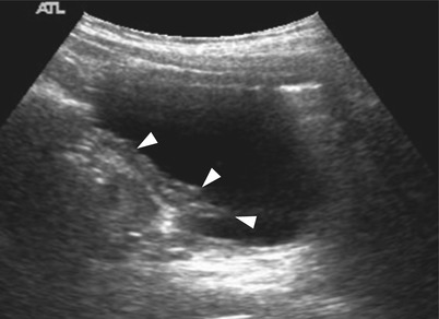
Fig. 26.1
Patent urachus in a 1-month-old neonate with urine leakage from umbilicus. (a) Longitudinal US shows a urine-filled patent urachus (arrowheads) extending from the dome of bladder (B) to the umbilicus (arrow). (b) Lateral view during a fistulography through the opening of umbilicus shows a fistulous tract (arrow) leading from the umbilicus to the dome of bladder
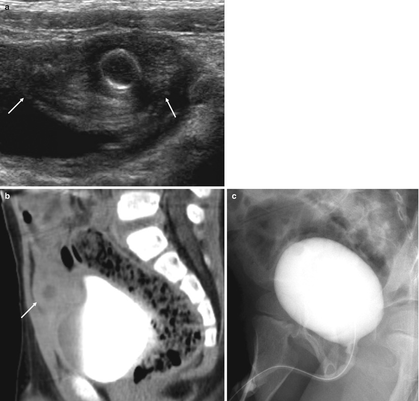
Fig. 26.2
Infected urachal cyst in a 3-year-old male. (a) Transverse US shows a complex cystic lesion (arrows) lying along the course of the urachus between the bladder and the umbilicus. The lesion demonstrates an internal cystic lesion with thicker outer wall. (b) Sagittal contrast-enhanced CT scan shows a peripheral enhancing, thick-walled cystic lesion (arrow) with perilesional infiltration at the bladder dome area. (c) Lateral view during a VCUG shows no evidence of urachal diverticulum
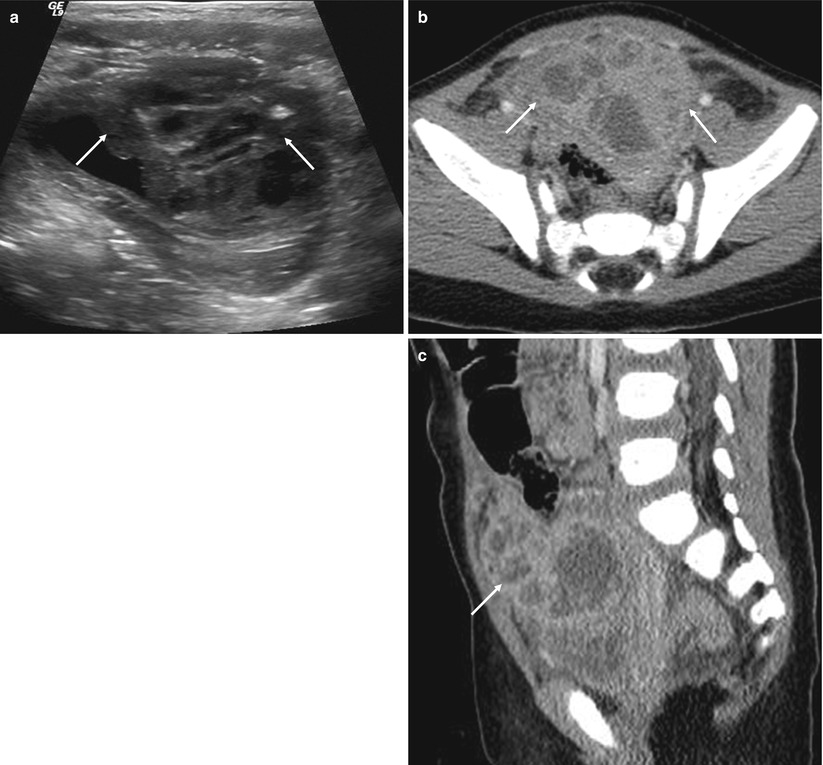
Fig. 26.3
Urachal abscess due to infected urachal cyst in a 1-year-old male. (a) Transverse midline US shows a large complex echoic mass (arrows) at the bladder dome area. (b, c) Axial (a) and sagittal (b) contrast-enhanced CT scans demonstrate enhancing thick-walled mass with low-density center consistent with abscess (arrows)
26.4.2 Bladder Exstrophy
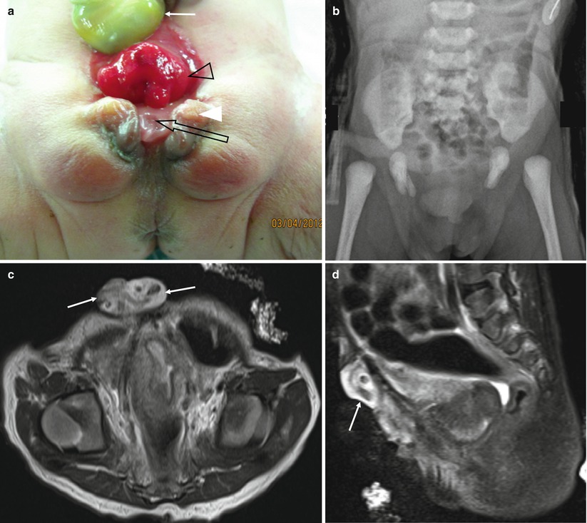
Fig. 26.4
Bladder exstrophy in a 2-day-old female infant. (a) Photograph shows the exposed bladder mucosa (open arrowhead) below the umbilical cord stump (white arrow). Also, note the exposed urethra (open arrow) and wide separation of labia (white arrowhead). (b) Anteroposterior supine radiograph shows wide separation of the symphysis pubis with outward rotation of the pelvic bone. The hips are dislocated. (c, d) Axial (c) and sagittal (d) T2-weighted MR images show a soft tissue outpouching (exposed bladder, arrows) below the umbilical cord stump. No bladder is noted in the pelvic cavity
26.4.3 Prune-Belly Syndrome
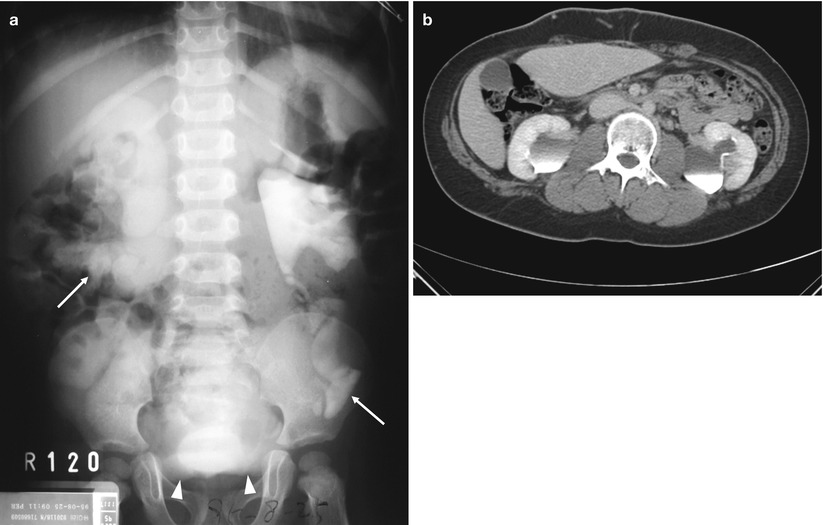
Fig. 26.5
Prune-belly syndrome in a 2-year-old male. (a) A 120-min intravenous urography (IVU) shows a huge bladder (arrowheads) and bilateral hydronephroureterosis with severe tortuous dilated ureters (arrows). (b) Fourteen years later, follow-up axial contrast-enhanced CT scans demonstrate absent or deficient abdominal musculature, hydronephrosis, and renal dysplasia with small kidneys
26.4.4 Bladder Diverticula
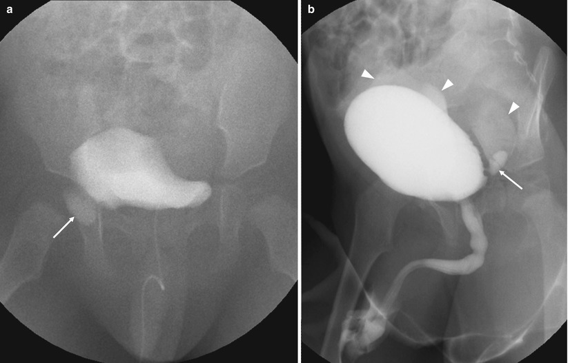
Fig. 26.6
Hutch diverticulum and bladder ear in a 6-month-old male. (a) VCUG shows a transient lateral herniation (arrow) of the bladder (“bladder ears”). (b) Right lateral oblique view during VCUG shows a small left-sided paraureteral bladder diverticulum (arrow) (“Hutch diverticulum”). Bilateral vesicoureteral reflux with severe tortuous ureters (arrowheads) is also seen
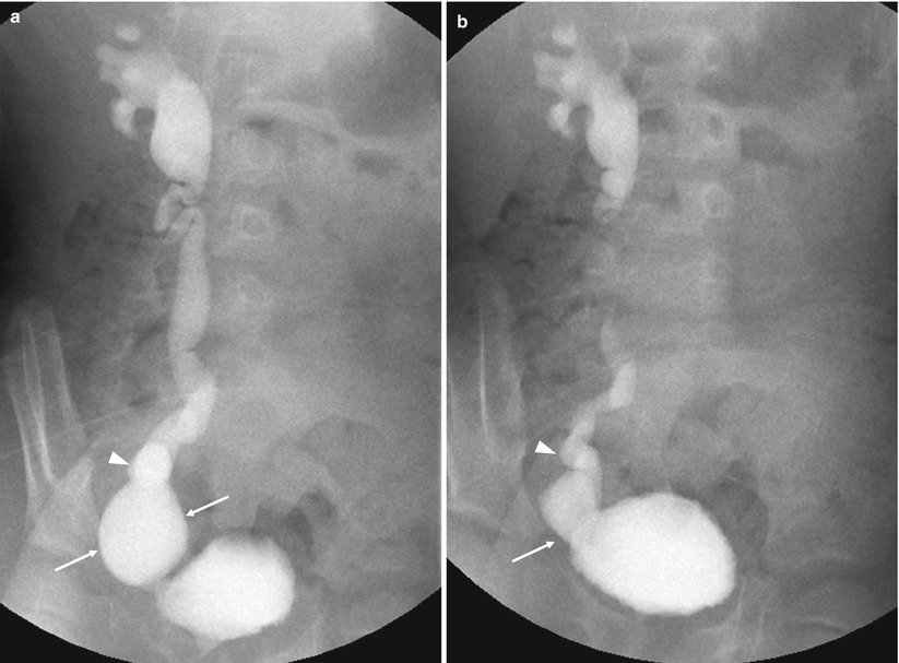
Fig. 26.7
Bladder diverticulum in a 3-year-old male. (a, b) Oblique (a) and anterior (b) images during VCUG show the ureter (arrowhead) inserting directly into the bladder diverticulum (arrows). This type of lesion prevents normal maturation of the ureterovesical junction and requires surgery to correct the reflux
26.4.5 Megacystis
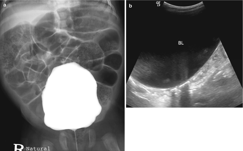
Fig. 26.8
Megacystis in a 29-month-old female. (a) Anterior image during VCUG shows a large urinary bladder. (b) Six years later, follow-up longitudinal US shows persistent large urinary bladder (BL)
26.4.6 Cystis
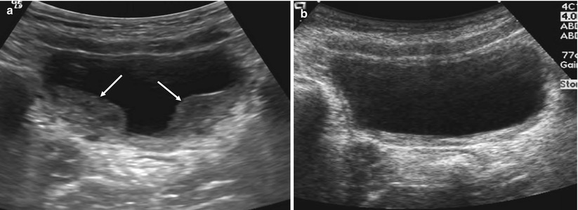
Fig. 26.9
Hemorrhagic cystitis in an 11-year-old male. (a) Transverse US shows focal thickenings of the posterior wall of the bladder (arrows). (b) Three weeks later, follow-up transverse US demonstrates a marked decreased wall thickening of the bladder
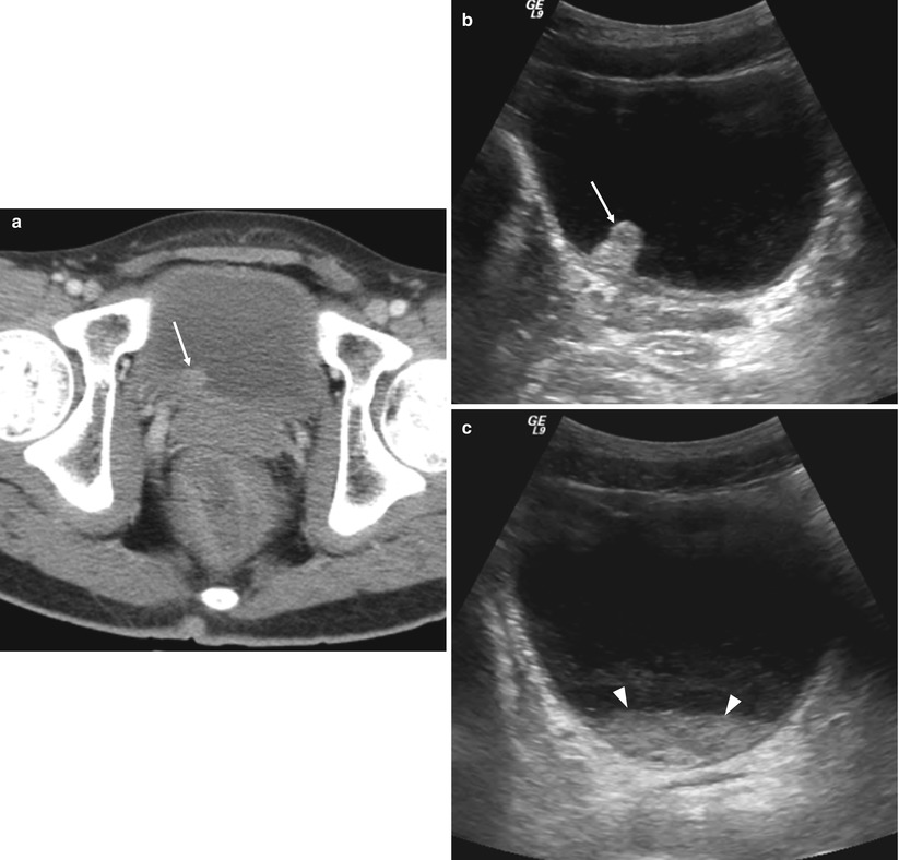
Fig. 26.10
Cyclophosphamide-induced hemorrhagic cystitis in a 14-year-old male who received allogeneic-peripheral stem cell transplantation (PBSCT) for aplastic anemia. (a) Axial contrast-enhanced CT shows focal protruding lesion (arrow) in the posterior wall of the bladder. (b, c) Transverse US images demonstrate a bilobed echogenic lesion (arrow in b) at the right ureterovesical junction level and an echogenic debris with fluid–fluid level (arrowheads in c) in the bladder base

Fig. 26.11
Cystitis cystica in an 11-year-old female. (a–d) Transverse US (a), VCUG (b), and contrast-enhanced CT images (c, d) show diffuse concentric wall thickening (arrows) at the bladder outlet. Bladder outlet is seen as a dumbbell shape with true bladder neck and false bladder neck on VCUG. Cystoscopic biopsy revealed cystitis cystica
26.4.7 Neoplasms of the Bladder
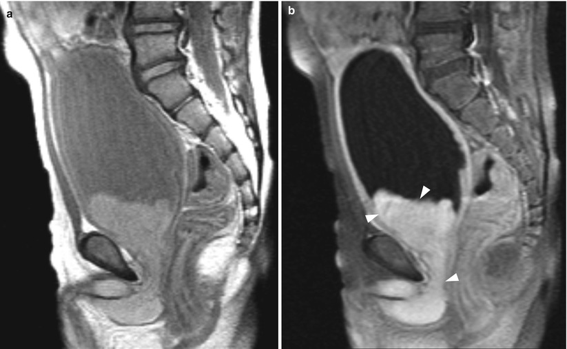
Fig. 26.12
Embryonal rhabdomyosarcoma in a 3-year-old male. (a, b) Sagittal T2-weighted (a) and contrast-enhanced T1-weighted (b) MR images show a lobulated mass (arrowheads) extending from the bladder base to the prostatic urethra
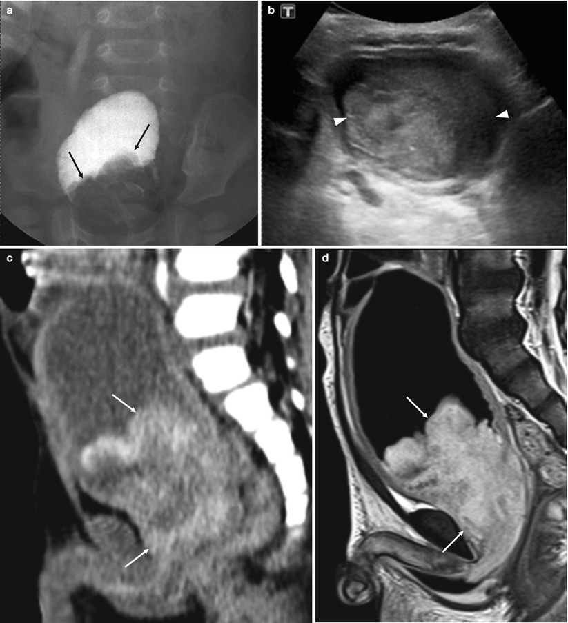
Fig. 26.13
Embryonal rhabdomyosarcoma in an 18-month-old male. (a) Bladder image from VCUG shows a lobulated filling defect (black arrows) in the bladder base. (b) Transverse US shows a solid intravesical mass (arrowheads) in the bladder base. (c, d) Sagittal contrast-enhanced CT (c) and T1-weighted MR (d) images show a large, heterogenous enhancing, polypoid mass (arrows) extending from the bladder base to the prostatic urethra and prostate
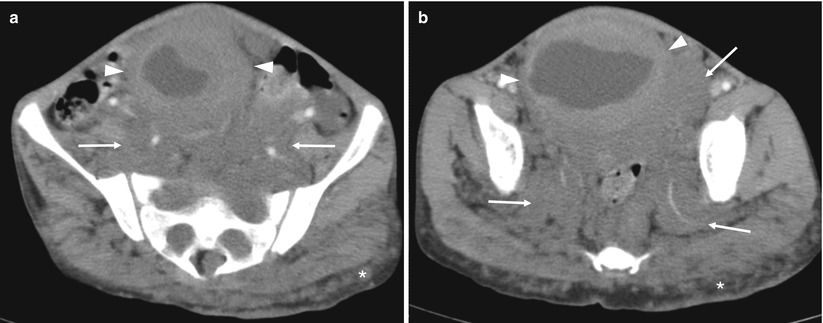
Fig. 26.14
Neurofibromatosis with direct involvement of the bladder in a 7-year-old female. (a, b) Axial contrast-enhanced CT scans show large peritoneal masses (arrows) with widening of sacral neural foramen and extending to the pelvis outlet through the sciatic notch. Also note multiple nodular masses (asterisks) extending to the subcutaneous layer of buttock. Bladder is displaced anteriorly and has diffuse wall thickening (arrowheads)