Chapter 11 Blood supply of the lumbar spine
The lumbar arteries
A pair of lumbar arteries arises from the back of the aorta in front of each of the upper four lumbar vertebrae.1,2 Occasionally, the arteries at a particular level may arise as a single common trunk which rapidly divides into right and left branches. At the L5 level, the fifth lumbar arteries arise from the median sacral artery but otherwise they resemble the other lumbar arteries.
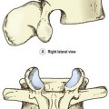
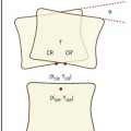
Each lumbar artery passes backwards around its related vertebral body (Fig. 11.1), lying in the concavity formed by the lateral surface of the vertebral body where it is covered by the tendinous arch of the psoas muscle. Upon reaching the level of the intervertebral foramen, the artery divides into several branches (Fig. 11.2).
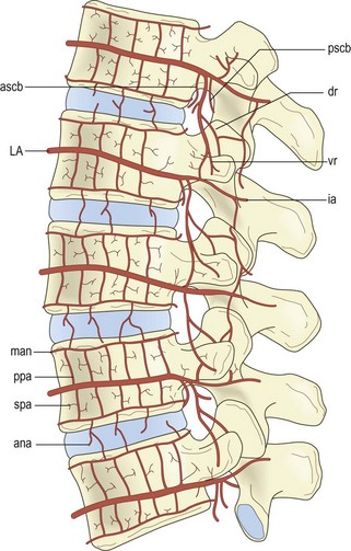
Lateral branches pass through the psoas and quadratus lumborum muscles eventually to supply the abdominal wall. Others pass with the ventral ramus and dorsal ramus of the spinal nerve supplying the paravertebral muscles innervated by these nerves. A substantial posteriorly directed branch passes below the transverse process, running perpendicular to the lateral border of the pars interarticularis of the lamina, to enter the back muscles (see Fig. 11.2).1,3 In addition to supplying the back muscles, the posterior branches of the lumbar arteries form anastomoses around the zygapophysial joints, which they supply, and plexuses that surround and supply the laminae and spinous processes.1
Opposite the intervertebral foramen, three medially directed branches arise from the lumbar artery (see Fig. 11.2). These are the anterior spinal canal branch, the posterior spinal canal branch and the radicular branch.1,3 The radicular branches are described in detail later.
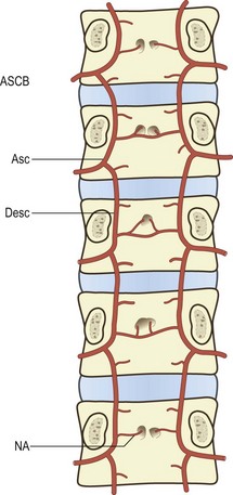
The anterior spinal canal branch at each level enters the intervertebral foramen and bifurcates into ascending and descending branches. The ascending branch crosses the intervertebral disc and circumvents the base of the pedicle above to anastomose with the descending branch from the next higher segmental level. In this way a series of arterial arcades is formed across the back of the lumbar vertebral bodies, i.e. along the floor of the vertebral canal (Fig. 11.3).
The lumbar veins
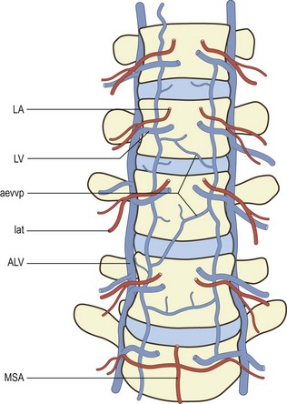
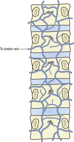
Several veins surround and drain the lumbar spine. These are the lumbar veins, the ascending lumbar veins and several vertebral venous plexuses. The lumbar veins accompany the lumbar arteries in their course around the vertebral bodies, and drain into the inferior vena cava (see Fig. 11.1). Opposite the intervertebral foramina the lumbar veins on each side communicate with the ascending lumbar vein, a long channel that runs in front of the bases of the transverse processes (Fig. 11.4). Inferiorly on each side, the ascending lumbar vein communicates with the common iliac vein while, superiorly, the right ascending lumbar vein joins the azygous vein, and the left ascending lumbar vein joins the hemiazygous vein.
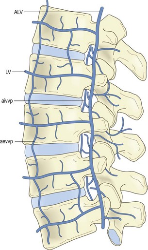
Over the anterolateral aspects of the lumbar spine, a variable series of vessels interconnect the lumbar veins to form the anterior external vertebral venous plexus (see Fig. 11.4). Within the vertebral canal, two other plexuses are formed. One covers the floor of the vertebral canal and is known as the anterior internal vertebral venous plexus (Fig. 11.5). The other lines the roof of the vertebral canal and is called the posterior internal vertebral venous plexus. Within the vertebral canal these plexuses extend superiorly to thoracic levels and inferiorly to sacral levels, and at each intervertebral foramen the two internal vertebral venous plexuses communicate with the ascending lumbar veins.
Blood supply of the vertebral bodies
As each lumbar artery crosses its vertebral body, it gives off some 10–20 ascending and descending branches called the primary periosteal arteries.2 Branches of these vessels supply the periosteum and outermost walls of the vertebral body (Figs. 11.2, 11.6
Stay updated, free articles. Join our Telegram channel

Full access? Get Clinical Tree


