• Well-defined, geographic signal abnormality, which tends to involve anterior 1/3 to 1/2 of vertebral body
• T2WI shows increased signal in multiple bodies, with abrupt transition to normal marrow signal
• T1WI C+ shows markedly diminished enhancement of affected vertebral body areas
No associated epidural or paravertebral soft tissue
Rare to involve posterior elements
Top Differential Diagnoses
• Infarction secondary to underlying systemic disease
Sickle cell
Acute leukemia (ALL or AML)
SLE
Lymphoma
Transplantation with graft vs. host disease
• Infarction secondary to aortic disease
Dissection
Abdominal aortic surgery
• Metastatic disease marrow infiltration
• Leukemia or lymphoma marrow tumor infiltration
• Granulomatous or fungal infection
Diagnostic Checklist
• Infarction as sign of systemic illness or malignancy, such as leukemia
• Associated with spinal cord infarction; useful as a confirmatory sign that nonspecific T2 hyperintensity in cord reflects infarction
(Left) Sagittal T1WI C+ MR in a patient with a new diagnosis of lymphocytic leukemia shows multiple well-defined, geographic foci of diminished enhancement in multiple vertebral bodies, with a rim of increased enhancement. There is no associated soft tissue mass and no disc involvement.
(Right) Axial T1WI C+ MR in a patient with multiple vertebral infarcts and a new diagnosis of ALL shows sharply marginated lesions in both right and left sides of the vertebral body with mild peripheral enhancement.
(Left) Sagittal T1WI MR shows multiple infarcts in a patient with ALL status post chemotherapy. Note well-defined low signal present in the anterior 1/2 of multiple vertebral bodies. (Courtesy M. Pathria, MD.)
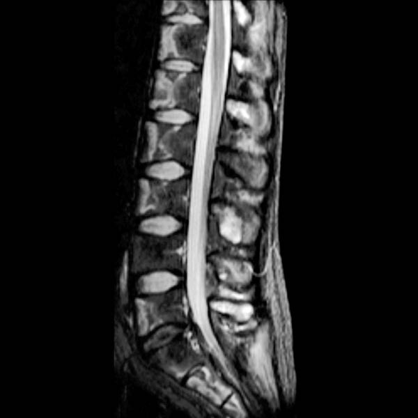
(Right) Sagittal T2WI FS MR in a patient with ALL status post chemotherapy shows focal, well-defined hyperintensity in the anterior 1/2 of multiple vertebral bodies, which also involves the sacrum. (Courtesy M. Pathria, MD.)
TERMINOLOGY
Synonyms
• Immature bone infarct, bone marrow infarction
Definitions
• Infarction of vertebral body cancellous bone and marrow secondary to systemic disease or aortic pathology
• Not osteonecrosis (Kümmell disease)
IMAGING
General Features
• Best diagnostic clue
Geographic T2 hyperintensity of multiple vertebral bodies with diminished enhancement in setting of systemic disease
Only gold members can continue reading. Log In or Register to continue


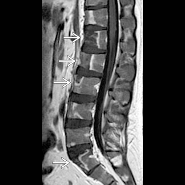
 in multiple vertebral bodies, with a rim of increased enhancement. There is no associated soft tissue mass and no disc involvement.
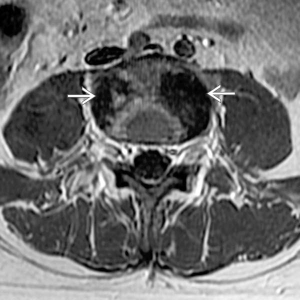
 with mild peripheral enhancement.
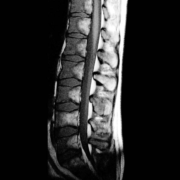
 Geographic T2 hyperintensity of multiple vertebral bodies with diminished enhancement in setting of systemic disease


