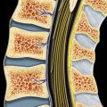
(Left) Axial graphic shows hematogenously disseminated lytic metastatic lesion to thoracic vertebral body and pedicle, with subsequent direct epidural tumor extension and cord compression.
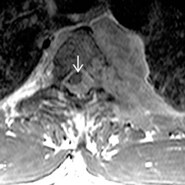
(Right) Axial T1WI C+ MR shows large hematogenously disseminated metastatic paravertebral mass lesion involving thoracic body, with direct tumor extension to posterior elements, chest wall, and epidural space with cord compression .
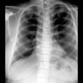
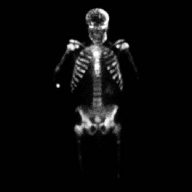
(Left) Bone scan in a case of extensive blastic prostate carcinoma shows multiple foci of increased radiotracer uptake consistent with diffuse bone metastasis.
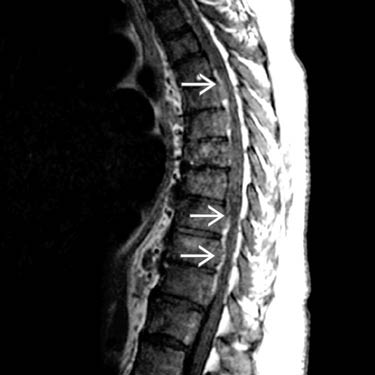
(Right) Sagittal T1WI C+ MR in a patient with extensive blastic prostate carcinoma shows diffuse abnormal decreased signal from all vertebral bodies and posterior elements with multiple foci of ventral epidural tumor extension , causing mild cord compression.
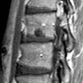
(Left) Sagittal T2WI MR in a patient with multiple myeloma shows 2 levels of bony tumor involvement and slight spinous process expansion. There is severe cord compression at both levels.
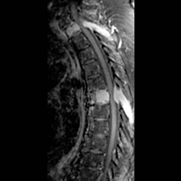
(Right) Sagittal T1 C+ FS MR of multiple myeloma shows 2 levels of focal tumor involvement with enhancement and slight spinous process expansion. Cord compression is present at both levels.
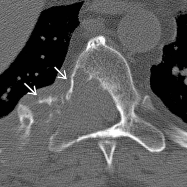
(Left) Axial NECT in a patient with renal cell metastasis shows mass destroying the right side of T7-T8 bodies, extending into posterior elements and crossing to involve right ribs and costovertebral joint. There is bony expansion with a thin rim “soap bubble” pattern .
Only gold members can continue reading. Log In or Register to continue
 .
.
 , causing mild cord compression.
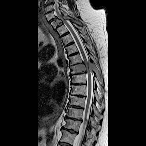
 .
.