• Osteopetrosis in differential diagnosis list for dense bones
• “Bone-within-bone” appearance not pathognomonic; may also be seen in young children during growth spurts
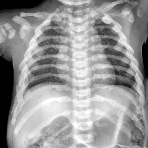
(Left) Anteroposterior radiograph of the thoracic spine and chest (infantile osteopetrosis) demonstrates marked diffuse sclerotic density within all bones. Note also abnormal thickened rib shape and multiple healing rib and clavicle fractures.
(Right) Lateral radiograph of the cervical spine (infantile osteopetrosis) reveals marked sclerotic bone density. Note also C2 pars fracture related to brittle bone with abnormal anterior angulation at C2/C3.
(Left) Sagittal T1WI MR (infantile osteopetrosis) shows universally abnormal low marrow signal intensity in all bones reflecting a combination of diffuse marrow replacement and sclerosis. The cerebellar tonsils are ectopic, probably acquired secondary to bone changes in the posterior fossa.
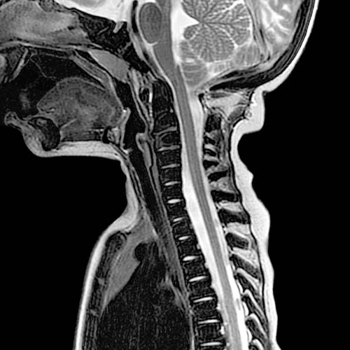
(Right) Sagittal T2WI MR (infantile osteopetrosis) reveals extensive abnormal low marrow signal intensity in all bones, including spine, skull base, sternum from marrow replacement, and osseous sclerosis.
TERMINOLOGY
Synonyms
• “Marble bone disease”
Definitions
• Heterogeneous grouping of hereditary osteoclast disorders, multiple autosomal dominant and recessive forms
IMAGING
General Features
• Best diagnostic clue
Diffuse increase in bone density
• Location
Entire skeleton
Radiographic Findings
• Radiography
Bowing deformity of bones, frequent fractures
Infantile form: Dense bones, marrow space obliteration
Delayed form: Thickened bone cortex, “bone-within-bone” appearance
CT Findings
• CECT
± extramedullary hematopoiesis
• Bone CT
Marked cortical thickening
MR Findings
• Sclerotic bone low signal intensity on T1WI, T2WI
• ± extramedullary hematopoiesis
Imaging Recommendations
• Best imaging tool
Radiography
Only gold members can continue reading. Log In or Register to continue


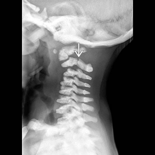
 related to brittle bone with abnormal anterior angulation at C2/C3.
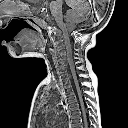
 are ectopic, probably acquired secondary to bone changes in the posterior fossa.