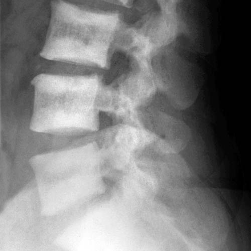
(Left) Lateral radiograph shows severe findings of renal osteodystrophy. There is marked sclerosis of the vertebral bodies adjacent to the endplate creating a striped appearance like a rugby jersey. The margins of the sclerosis are poorly defined, in distinction to the sharply demarcated sclerotic regions seen in osteopetrosis.
(Right) Lateral radiograph shows fairly mild changes of renal osteodystrophy (OD). Ill-defined sclerosis is present adjacent to the endplates, and bony trabeculae are blurred.
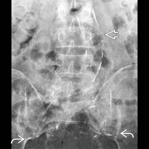
(Left) Anteroposterior radiograph in the same patient shows sacroiliac erosions due to 2° hyperparathyroidism. Accelerated arterial calcifications are due to the abnormal calcium/phosphate levels.
(Right) Axial bone CT through the thoracic spine shows sclerosis and blurred trabeculae due to renal osteodystrophy. The appearance is more uniform and diffuse than that of sclerotic metastases.
TERMINOLOGY
Abbreviations
• Renal osteodystrophy (OD)
Definitions
• Chronic kidney disease-mineral and bone disorder (CKD-MBD): Constellation of abnormalities seen in progressive kidney disease, including metabolic abnormalities, disturbances in bone modeling and remodeling, and extraskeletal calcification in arteries and soft tissues
• Renal osteodystrophy (OD): Only defines bone pathology associated with CKD
IMAGING
General Features
• Best diagnostic clue
Rugger jersey spine
• Location
Involves axial and appendicular skeleton
Radiographic Findings
• Radiography
Osteosclerosis &/or osteopenia
Only gold members can continue reading. Log In or Register to continue


 due to 2° hyperparathyroidism. Accelerated arterial calcifications
  are due to the abnormal calcium/phosphate levels.
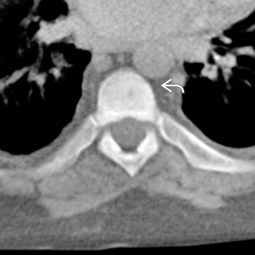
 and blurred trabeculae due to renal osteodystrophy. The appearance is more uniform and diffuse than that of sclerotic metastases.


