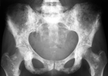
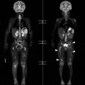
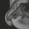
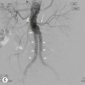
A. Mark Davies, Steven L.J. James, Asif Saifuddin Bone tumours can be generally divided into two groups: benign, including tumour-like lesions (see Chapter 47), and malignant. The malignant category can be subclassified into primary, secondary and metastatic. In common parlance the terms secondary and metastatic are frequently used interchangeably. In the context of this chapter, however, the term secondary is reserved for those tumours arising from malignant transformation of a pre-existing benign bone condition. Bone metastasis or metastatic disease refers to the spread of a malignant tumour from its primary site to a non-adjacent part of the skeletal system. They are relatively common, frequently found on imaging to be multiple and are the commonest bone malignancy over 40 years of age. Approximately, 10% of carcinoma metastases will present as a solitary bone lesion. A solitary lesion in a middle-aged or elderly patient is, therefore, still more likely to be a metastasis than a primary malignant bone tumour. Post-mortem studies have shown that bone is the third commonest site of metastatic spread after the lung and liver. Metastases are assumed to arise from venous tumour emboli, from the primary tumour or from prior disease spread to the regional lymph nodes or other metastases, e.g. in the lung. Venous embolisation of malignant cells is multifactorial but is particularly related to the vascularity of the primary tumour and/or access to a valveless venous plexus, e.g. Batson’s vertebral plexus. Bone metastasis is a relatively late occurrence in the natural history of malignant disease because the lungs trap most tumour emboli. It is recognised that bone metastases may occur without obvious evidence of pulmonary involvement due to: 1. Pulmonary lesions that are present but occult; 2. Transpulmonary spread of malignant cells; 3. Paradoxical embolisation via a patent foremen ovale; and 4. Retrograde venous embolism with involvement of the vertebral column. Arguably, the incidence of truly occult pulmonary metastases is less these days due to the superior sensitivity of multidetector CT of the chest routinely used in cancer staging as compared with standard radiography or single slice CT employed in the past. The most common primary malignancies metastasising to bone are carcinoma of the breast, bronchus, prostate, kidney and thyroid, making up over 80% of all patients with bone metastases. The overall incidence of bone metastases during life is uncertain and will depend on the sensitivity of the imaging technique employed. At the time of death the prevalence of bone metastases is approximately 80–85% for breast and prostate cancer, more variable for lung cancer with a quoted range of between 40 and 80%, 50–60% for thyroid cancer and 20–35% for renal cancer. With modern improved cancer management leading to longer survival times, bone metastases have become a fairly common occurrence in everyday medical practice. The commonest sites of metastatic involvement are those containing red bone marrow, explaining why the axial skeleton is affected more commonly than the appendicular skeleton in adults; hence, the predilection for the spine, pelvis, proximal femora and humeri, ribs, sternum and skull (Fig. 48-1). Spinal metastases occur most commonly in the thoracic vertebrae, but carcinomas arising within the pelvis, particularly prostatic carcinoma, show a predilection for the lumbosacral spine. Unexplained back or limb pain in a patient with a history of carcinoma may indicate a skeletal metastasis. Non-mechanical pain (i.e. bone pain at rest) is highly suggestive of tumour, be it primary or metastatic. A metastasis may present with a pathological fracture following minor trauma. Radiographs are necessary to reveal the pre-existing lesion. Elevation of the serum alkaline phosphatase is a non-specific finding and typically is not seen until multiple bone metastases have developed. Bone metastases may be lytic (most common) (Fig. 48-2), mixed lytic and sclerotic and predominantly sclerotic (Fig. 48-3). Small lytic lesions confined to the medullary bone can be difficult to identify on radiographs, particularly in complex anatomical areas such as the pelvis and spine or where there is reduced bone density (osteopenia). It is much easier to detect a metastasis as and when there is evidence of cortical destruction. It is for this reason that vertebral metastases are frequently first detected on radiographs as showing destruction of the cortex of a pedicle even though cross-sectional imaging may reveal fairly extensive destruction of the medullary bone in the adjacent vertebral body (Fig. 48-4). As the destructive process increases, a soft-tissue mass may develop. A periosteal reaction may be seen but this is usually less pronounced than those seen in primary bone malignancies. Exceptions include some prostatic metastases and mucinous adenocarcinoma metastases from the colon. Some carcinomas, such as renal, almost always produce lytic metastases. Others, such as prostate, are most commonly osoteoblastic, while breast metastases may show a mixed appearance. It should be noted that the osteoblastic reaction reflects the response of the host bone to the metastasis rather than the production of tumour bone, which is a characteristic of osteosarcoma. Cortical-based metastases are less common than medullary and tend to arise along the diaphyses of the long bones from lung, kidney and breast primaries (Fig. 48-5). The importance of identifying multiple lesions cannot be overemphasised, as an individual metastasis may be indistinguishable from a primary malignant bone tumour. For this purpose, 99mTc-MDP bone scintigraphy is the most cost-effective method, although both whole-body MRI and 18F-fluoride PET/CT are considered more sensitive. A variety of scintigraphic abnormalities are seen, with the most common being multiple foci of increased skeletal activity, predominantly in the axial skeleton (Fig. 48-1). Abnormal increased activity relies on an intact local blood supply and increased host bone osteoblastic activity. Metastases that have infarcted or stimulate no host osteoblastic response may appear as photopenic areas or ‘cold spots’, most commonly seen with larger renal metastases. Occasionally a combination of ‘hot’ and ‘cold’ lesions occurs. Diffuse osteoblastic metastatic disease, typically from breast or prostate carcinoma, may result in a ‘superscan’ appearance where there is generalised increase in skeletal activity with a relative paucity of renal activity. It is important to recognise that not all foci of abnormally increased activity on bone scintigraphy, in a patient being investigated for suspected metastatic disease, need necessarily be due to metastases. Foci of activity in the ribs, particularly if contiguous, suggest old fractures, linear foci peripherally in the sacrum suggest insufficiency fractures and intense expanded foci of activity extending up to a joint margin may indicate Paget’s disease. Any area of unexplained abnormal uptake should be correlated with further up-to-date imaging. Generally, MRI is utilised as it is more sensitive than radiography, and more specific than scintigraphy. Most metastases are located within the medulla and show reduced signal intensity (SI) on T1-weighted sequences, with increased SI on T2-weighted or fat-suppressed sequences. The identification of a hyperintense ‘halo’ around a lesion is a feature highly suggestive of a metastasis on fluid-sensitive images. Both whole-body MRI and FDG-PET are being increasingly utilised in the detection and monitoring of bone metastases.1,2 The efficacy of PET can be increased with fused anatomical imaging, e.g. PET/CT.3 Prostatic metastases are typically osteoblastic (sclerotic) (Fig. 48-6) or mixed, with purely lytic lesions being very rare. Occasionally, long bone metastases may exhibit a prominent ‘sunburst’ periosteal reaction mimicking an osteosarcoma, a rare disease in this age group in the absence of Paget’s disease or previous radiotherapy. Disseminated prostatic metastases may produce confluent sclerosis on radiographs, simulating other disorders such as myelofibrosis and Paget’s disease, although the latter condition can usually be excluded by noting the absence of bony expansion. Assessment of the prostate serum antigen (PSA) level can be useful in determining the value of performing bone scintigraphy. If the PSA level is <10 ng/mL the likelihood of positive bone scintigraphy is <1%. This increases to approximately 10% if the PSA level is 10–50 ng/mL and 50% with a PSA level >50 ng/mL. While 99mTc-MDP bone scintigraphy may be adequate for the initial identification of prostatic metastases, FDG-PET is preferred after treatment for distinguishing persistent active metastases from healing bone. Most breast bone metastases are lytic (Fig. 48-2) but breast carcinoma is the commonest cause of osteoblastic metastases in women (Fig. 48-3). About 10% of metastases are purely osteoblastic and 10% mixed. Diffuse marrow infiltration may occur and only become radiographically visible when sclerosis develops after therapy. In this situation the sclerosis may be mistaken for disease progression rather than a response to treatment. Breast metastases have a predilection for the spine, pelvis and ribs. Patients may present with acute-onset vertebral collapse at one or more levels and positive neurology. In this clinical situation the differential diagnosis would include benign causes such as osteoporotic collapse and malignant causes such as metastases from other primaries, lymphoma and myeloma. Further investigation with MRI ± diffusion-weighted imaging would be mandatory. Bone scintigraphy is usually sufficient to confirm/exclude bone metastases when clinical or laboratory parameters are suspicious but after treatment, follow-up studies may show increased activity due to the ‘flare phenomenon’ secondary to the normal healing response of bone. Again, in this context FDG-PET may be preferred with increasing activity indicating a lack of response to treatment. Lung metastasis to bone is common. Most spread to the axial skeleton and are typically lytic. Metastases to the hands and feet (acrometastases), although rare, originate from a lung primary in approximately half of cases (Fig. 48-7).4 Bronchogenic carcinoma is the commonest source of cortical-based metastases with large lesions showing the so-called ‘cookie-bite’ appearance (Fig. 48-5). It is also the commonest disorder associated with hypertrophic osteoarthropathy, formerly known as hypertrophic pulmonary osteoarthropathy. A single or multiple layer periosteal reaction along the distal long bones, including the hands and feet, characterises this condition. Bone scintigraphy will reveal symmetrical increased linear activity along the long bones, known as ‘double stripe’ or ‘parallel tract’ sign. Renal carcinoma is the commonest primary malignancy associated with solitary bone metastases. Solitary or multiple lesions are invariably lytic and may be expansile (blowout) and trabeculated. So-called expansile metastases may also be typically seen in thyroid metastases (Fig. 48-8) and less commonly in breast and lung. A similar radiographic appearance may be seen with plasmacytoma and, if subarticular, in a younger age group, giant cell tumour of bone. Renal metastases tend to be hypervascular, exhibiting multiple serpiginous signal voids within and around the periphery of the lesion on MRI.5 After extension to locoregional lymph nodes, malignant melanoma can spread to almost any part of the body, including the axial skeleton. It is important to appreciate that melanoma metastases may be clinically silent and that there can be a long latent period between treatment of the primary lesion and the development of the metastasis. Most bone metastases from melanoma are lytic. Melanin is paramagnetic and, depending on the concentration, the metastasis may show mild hyperintensity on T1-weighted MR images. When a known primary malignancy is established, the presence or absence of metastases is part of the routine surgical staging process. This will vary, depending on the nature of the primary and local/national guidelines. In most situations bone scintigraphy is adequate for assessing the whole skeleton (Fig. 48-1). Combining the study with SPECT may increase the sensitivity in detecting small lesions in anatomically complex areas such as the spine. Whole-body MRI may be preferred because of its increased sensitivity but it should be noted that if only coronal sequences are obtained both rib and spinal lesions may be overlooked. When there is no history of prior malignancy then, in patients over 40 years of age, a CT study of the chest, abdomen and pelvis should be performed. Should the staging studies show the bone lesion to be solitary, then an image-guided needle biopsy will be required, irrespective of whether there is a documented prior history or current imaging evidence of a primary malignancy elsewhere. This is because the bone lesion need not necessarily be related to that other tumour and failure to establish the correct diagnosis could lead to inappropriate management prejudicial to the long-term prognosis for the patient. This biopsy should be performed so as to avoid contamination of uninvolved anatomical compartments in case the histology reveals a primary malignancy of bone rather than a metastasis. In cases with disseminated metastases to multiple organ systems, where palliative care is the only reasonable management, needle biopsy is arguably unnecessary. In patients with a suspected primary malignancy and bone metastases, a needle biopsy of the most accessible bone lesion may be the quickest and easiest way to establish a definitive tissue diagnosis. An important cause of morbidity in patients with bone metastases is the development of pathological fractures. This is most common in the spine, with or without neurological compromise, and in the lower limbs because of weight-bearing. It is clearly advantageous for the patient if these debilitating fractures can be prevented with prophylactic fixation. In the long bones the Mirels’ scoring system is widely employed to assess fracture risk (Table 48-1).6 TABLE 48-1 Mirels’ Rating System for Prediction of Pathological Fracture Risk of Metastases in Long Bones as Measured on Radiographs6 a Relative proportion of bone width involved with tumour. b Non-perithrochanteric lower extremity. c Mixed lytic and blastic. In the past the identification of bone metastases usually meant an early demise for the patient; but nowadays modern therapies and improved surgical techniques are leading to longer survival times. Therefore, follow-up imaging to assess response of bone metastases to treatments is not uncommon. Such imaging can be difficult to interpret: while disease progression is usually obvious, it can be more problematic to confirm lack of progression. The ‘flare phenomenon’ on bone scintigraphy is an example where a good response to therapy may be misinterpreted as disease progression. It is also important to recognise that new bone lesions may not be further metastases but actually caused by the drugs administered. This includes bone infarction after chemotherapy and/or steroids and necrosis of the mandible and atypical stress fractures in the femora after bisphosphonate therapy.7 It is likely that FDG-PET will have an increasing role in the assessment of the response of bone metastases to therapy. This is a distinctly less common occurrence than in adults. In the younger child the commonest disseminated neoplasms affecting bone are neuroblastoma and leukaemia, which may have identical radiographic appearances. The commonest paediatric soft-tissue sarcoma and also the commonest to metastasise to bone is rhabdomyosarcoma, with a 10% incidence identified at time of initial diagnosis. Retinoblastoma may involve bone by direct spread and blood-borne metastases. It is also one of a number of conditions associated with an increased incidence of osteosarcoma. Both osteosarcoma and Ewing sarcoma may present or develop local (skip) or distant bone metastases. The commonest intracranial neoplasm to metastasise to bone outside the skull is cerebellar medulloblastoma, typically after surgery to the primary tumour.
Bone Tumours (2)
Malignant Bone Tumours
Introduction
Bone Metastases
Distribution of Bone Metastases
Diagnosis of Bone Metastases
Clinical
Radiological Features
Prostate.
Breast.
Lung.
Kidney.
Melanoma.
Radiological Investigation of Bone Metastases
Score
Site
Nature
Sizea
Pain
1
Upper extremity
Blastic
< 
Mild
2
Lower extremityb
Mixedc
 –
–
Moderate
3
Peritrochanteric
Lytic
> 
Severe

Bone Metastases in Children
Primary Malignant Neoplasms of Bone
Primary malignant tumours of the skeleton are rare, accounting for only 0.2% of all neoplasms, with an annual incidence of new diagnoses of approximately 1/100,000 population. Major advances in the past 40 years in chemotherapy and limb-salvage surgery have vastly improved the outcome, such that in specialist centres, the 5-year survival for appendicular osteosarcoma now exceeds 60%. The classification of these tumours, as described by the World Health Organisation, is based on their histopathological characteristics.8 While the tumours themselves have remained unchanged over the years, the nomenclature has metamorphosed as successive generations of pathologists have attempted to refine and reclassify these disorders.
Radiographs remain of fundamental importance in the detection and diagnosis of a primary bone tumour, be it benign or malignant. Radiographic features suggestive of an aggressive and therefore more likely to be malignant lesion include a wide zone of transition (permeative or moth-eaten), an aggressive periosteal reaction (onion-skin, Codman angle or spiculated), cortical destruction and a soft-tissue mass.9 Matrix mineralisation does not help to distinguish benign from malignant disorders but can be an indicator of the underlying histopathological origin, be it osteoid (i.e. bone-forming) or chondroid (i.e. cartilage-forming).
Chondroid Origin
Chondrosarcoma
Chondrosarcoma is a malignant cartilage-forming tumour accounting for 8–17% of all biopsied primary bone tumours and 20–25% of all biopsied malignant bone tumours. It is classified as central if arising within medullary bone (Fig. 48-9) and peripheral if arising from the surface of bone. It is further subclassified as primary if arising de novo or secondary if arising from a pre-existing bony lesion, usually an enchondroma (central type) or osteochondroma (peripheral type) either as a solitary occurrence or as part of the bone dysplasias of Ollier’s disease (multiple enchondromatosis), Maffucci’s syndrome (multiple enchondromatosis and soft-tissue angiomas) and hereditary multiple exostoses (diaphyseal aclasis) (Fig. 48-10). The incidence of malignant change in solitary enchondroma and osteochondroma is <1% and in hereditary multiple exostoses <5%. The incidence is somewhat higher, at approximately 20% in Ollier’s disease and 30% in Maffucci’s syndrome.
Chondrosarcoma is also classified according to its histological grade. These are defined as low grade (grade 1; 45–50% of cases), intermediate grade (grade 2; 30–40% of cases), high grade (grade 3; 10–25% of cases) and dedifferentiated, which refers to the development of areas of high-grade non-cartilaginous sarcoma within the chondrosarcoma (10% of cases), typically osteosarcoma or spindle cell sarcoma (Fig. 48-11).10 Other distinct variants include mesenchymal and clear-cell chondrosarcoma.


