Bones
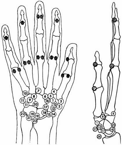 Figure 63-1. Accessory ossicles of the hand. The shaded circles indicate locations of sesamoid bones. 1. Epitrapezium 2. Calcification (bursa, flexor carpi radialis) 3. Paratrapezium (petrapezium) 4. Trapezium secundarium 5. Trapezoides secundarium 6. Os styloideum 7. Ossiculum Gruberi 8. Capitatum secundarium 9. Os hamuli proprium 10. Os vesalianum 11. Os ulnare externum (calcified bursa or tendon) 12. Os radiale externum 13. Fissure of traumatic origin 14. Persisting ossification center of the radial styloid process 15. Intercalary bone between the navicular and the radius (paranavicular) 16. Os carpi centrale 17. Hypolunatum 18. Epilunatum 19. Accessory bone between the lunate and the triangular bone 20. Epipyramis 21. So-called “os triangulare” 22. Persisting center of the ulnar styloid 23. Small ossicle at the level of the radioulnar joint 24. None 25. Avulsion from the triangular bone; no accessory ossicle 26. Tendon or bursal calcification 27. Calcification of the pisiform (Keats TE. Atlas of normal Roentgen variants that may Simulate disease, 5th ed. St. Louis: Mosby-Year Book, 1992:420-430. Fig. 6-139, p. 430). |
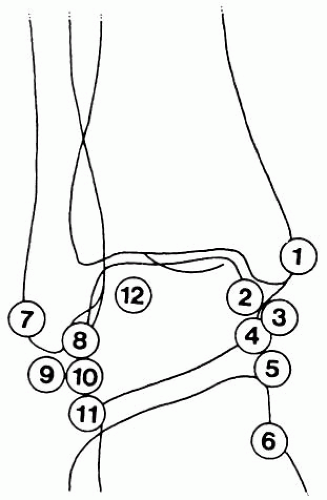 Figure 63-2. Accessory ossicles of the ankle. 1. Accompanying shadow on the internal malleolus (patella malleoli) 2. Intercalary bone (or sesamoid) between the internal malleolus and the talus 3. Os subtibiale 4. Talus accessorius 5. Os sustentaculi 6. Os tibiale externum 7. Os retinaculi 8. Intercalary bone (or sesamoid) between the external malleolus and the talus 9. Os subfibulare 10. Talus secundarius 11. Os trochleare calcanei 12. Os trigonum
Related posts:Stay updated, free articles. Join our Telegram channel
Full access? Get Clinical Tree
 Get Clinical Tree app for offline access

|