Motor or sensory deficits, ataxia, diplopia, dysarthria, dysmetria, vertigo, visual field deficit, cranial nerve dysfunction, syncope
• Treatment
Brace to restrict head motion, surgical fusion to prevent atlantoaxial rotation, vertebral artery decompression
Vertebral artery stenting
Diagnostic Checklist
• Temporary positional occlusion of 1 vertebral artery during course of daily activities may be normal, if asymptomatic
• Hypoplasia or stenosis of contralateral vertebral artery predisposes patients to vertebrobasilar ischemic attacks during head rotation
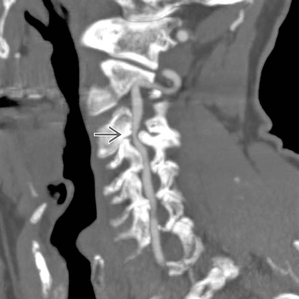
(Left) Coronal oblique CTA shows focal extrinsic narrowing of the left vertebral artery at the C3-C4 level due to facet degenerative hypertrophy and osteophyte formation .
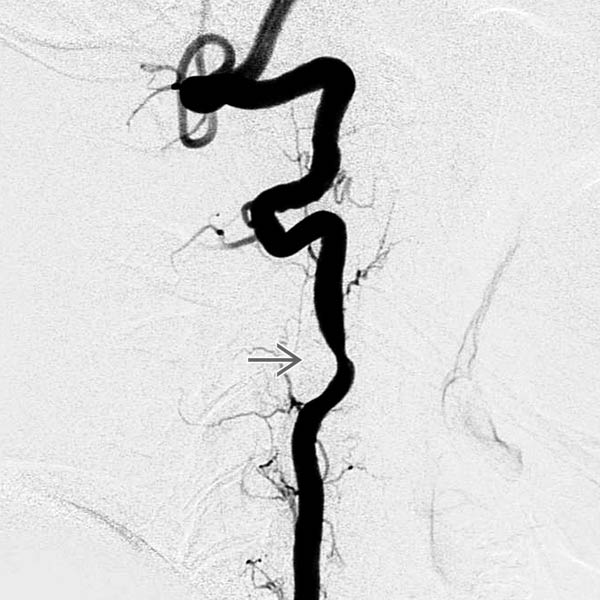
(Right) Lateral catheter angiography in a neutral position shows moderate focal stenosis at the C3-C4 level due to osteophytic compression .
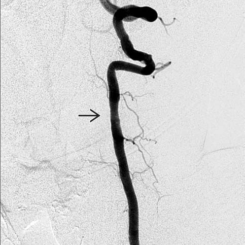
(Left) Anteroposterior catheter angiography with the head turned to the right (asymptomatic head turn side) shows patent left vertebral with mild C3-C4 level narrowing .
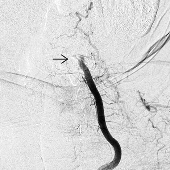
(Right) Anteroposterior catheter angiography with the head turned to the left (symptomatic head turn side) shows occlusion of the vertebral artery at the level of the osteophytic compression .
TERMINOLOGY
Synonyms
• Bow hunter stroke, positional occlusion of vertebral artery, rotational occlusion of vertebral artery
Definitions
• Vertebrobasilar insufficiency secondary to mechanical occlusion or stenosis of vertebral artery during head rotation due to fibrous band or bony prominence
IMAGING
General Features
• Best diagnostic clue
Occlusion or stenosis of vessel with head positional dependence based on ultrasound, MRA, CTA, or catheter angiography
• Location
Along course of vertebral artery, typically at C1-C2 junction
• Morphology
Stenosis or occlusion of vertebral artery with head position change
CT Findings
• CTA
Stenosis or occlusion of vertebral artery with head turned to symptom-producing side
Only gold members can continue reading. Log In or Register to continue
 Brace to restrict head motion, surgical fusion to prevent atlantoaxial rotation, vertebral artery decompression
 .
.
 .
.
 .
.
 .
.


