Fig. 29.1
(a–e) Simple small bowel obstruction. (a) CT scout view shows overdistention of small bowel loops with multiple and thin valvulae conniventes in the left abdominal quadrants. (b) Contrast-enhanced axial CT scan shows dilated jejunal loops with multiple and thin valvulae conniventes and air-fluid levels. (c) Contrast-enhanced axial CT scan illustrates the site of small bowel obstruction “transition zone” (arrows). (d) Same scan as (c) demonstrating the site of obstruction (red line) and the dilated loop with the “beak” (yellow line) at the “transition zone.” Note absence of fluid in the peritoneal cavity. The vascular enhancement of the obstructed loops and their vascular supply are preserved (e)
29.1.1.2 Decompensated SBO
Usually, the gastrointestinal tract handles 8–9 l of fluid every day, most of which is reabsorbed by the small bowel. If the occlusive status persists, simple SBO develops into decompensated SBO. The increasing intraluminal tension causes a change of parietal microcirculation, which hampers the ability of the bowel. This happens only when the intraluminal pressure is greater than the pressure in the capillary vessels, causing an alteration of vascular permeability. The bowel loop, progressively, becomes decompensated. A definite flow of fluids forms out of the intestinal wall, into the lumen, and in the peritoneal cavity. Nevertheless, the wall does not become thicker, because the tension of the lumen squeezes it like a sponge. The stretched and squashed wall becomes thin and tight.
IV contrast-enhanced CT shows thin walls with preserved and homogeneous CE.
The wall transudates liquid into and out of the loop; the transudate appears in the peritoneal cavity. As in all states of imbalance, the decompensated bowel may be acute or chronic, and it may resolve or deteriorate. In simple and decompensated SBO, the mortality is about 3 %.
Simple and decompensated SBO (Fig. 29.2) may resolve after medical therapy and placement of a nasogastric tube.
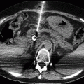
Fig. 29.2
(a–d) Decompensated small bowel obstruction. (a) Anteroposterior supine radiograph shows distended small bowel loops with multiple and thin valvulae conniventes. (b) Contrast-enhanced axial MDCT scan confirms dilated small bowel loops with mixed stasis and a small amount of fluid between the loops. The transudate collects and is visible in the pelvic recesses (c); the distal ileal loops are collapsed. (d) Maximum intensity projection image demonstrates all the above-mentioned signs and, furthermore, the “transition zone” between dilated and collapsed loops (circle). The mesenteric vessels and their adipose sheet are normotransparent. At surgery an adhesional band was found
29.1.1.3 Complicated SBO
Complicated SBO means an occlusive state complicated by vascular changes of the bowel wall. These complications may present with two different modalities:
Vascular changes due to reaction
Vascular changes due to strangling-strangulation
Regardless of the type, in the initial phase vascular change in the loop is a reversible phenomenon with recovery of the intestinal vitality and function still possible. Nevertheless, without a timely resolution, the final condition of this acute secondary venous ischemia is necrosis, gangrene, perforation, and peritonitis.
29.1.1.4 Vascular Changes due to Reaction
In the progression of SBO, vascular suffering due to reaction may appear. The origin of this event is multifactorial and unpredictable.
Main factors include:
The obstructive mechanism, duration of occlusion, and onset of complex mechanisms
The mesenteric and enteric circulation, already uncertain because of diffuse atherosclerotic disease
The pathological remains that alter the abdominal habitat
The general status and age of the patient
IV contrast-enhanced CT shows high contrast enhancement in the wall with normal or borderline thickness. The continuous vascular afflux causes repeated engorgement and a vascular dilation with intramural congestion: the wall becomes thick.
Usually, a reactive loop is near the obstructive site. The venous congestion of the loop may cause passage of blood into the peritoneal cavity. Mesenteric involvement is absent or minimal.
29.1.1.5 Vascular Changes due to Strangulation
The mechanism of strangulation involves the loop and its mesentery. In these cases, the perfusion deficit involves the venous circulation which is more easily compressible and collapsible. Impairment of the arterial circulation is very late. The obstacle to the blood outflow causes:
Intramural venous congestion with pathological wall thickening of the loop.
Hemorrhagic engorgement of the mesentery. The swollen and edematous mesentery curves the loops anteriorly, spreading them out. There is prominent vasculature with numerous dilated vessels.
Early appearance of peritoneal fluid. The intraperitoneal fluid will be initially found in the recesses between the loops, then in the peritoneal recesses of the mesentery, and finally free in the peritoneal cavity.
Strangulation has been observed in 10–15 % of patients who underwent surgery for SBO. Frager and Baer (1995) report a mortality of about 8 % if the surgical intervention is performed within 36 h from the beginning of symptoms. Over 36 h, the mortality increases to about 25 %.
29.1.1.6 Volvulus
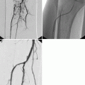
The term volvulus derives from the Latin verb “volvere” – to roll or twist – and defines loop torsion on its mesenteric axis; the mesentery wraps itself into coils (Fig. 29.3). This mechanism causes abnormal location of loops and vessels, which appear to converge toward the torsive point. Loops assume a spiral configuration. Vessels are stretched and twisted until the artery and vein are inverted.
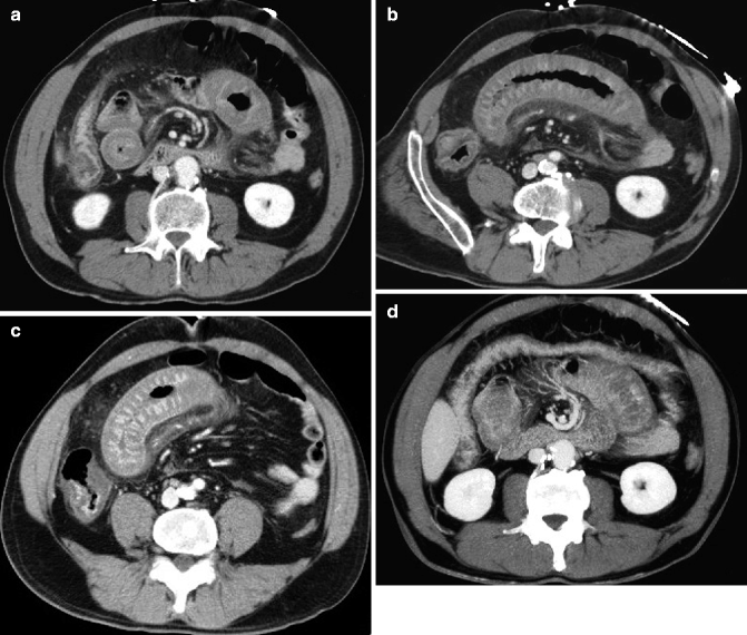
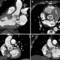
Fig. 29.3
(a–d) Small bowel volvulus. (a–c) Contrast-enhanced axial MDCT scans show ileal loops with thickened submucosa and slight reduced wall enhancement. Opaque inhomogeneous adipose tissue of the adjacent mesentery with loss of transparency of the perivascular adipose sheet. Note also the twisted mesenteric vessels: “whirl sign” in (a). (d) Maximum intensity projection image highlights the “whirl” of the superior mesenteric vein around the artery and the engorgement of the mesenteric vessels
Volvulus may spontaneously resolve. Formation of volvulus does not necessarily imply strangulation of the feeding vessel pedicle. This ultimate event verifies only in cases of a particularly serrated strangulation.
29.1.1.7 Closed Loop
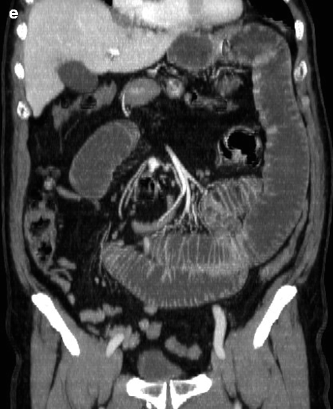
Closed loop means an obstructed loop at two adjacent points along its course (Fig. 29.4). Formation mechanism of closed loop is a single obstructed focus which interacts on two points of the same intestinal tract. Numerous formation mechanisms of SBO may generate closed loops. Actually, it may be caused by an adhesive band, entrapped-incarcerated hernia or laparocele, or also volvulus. Warning: evidence of a closed loop is not synonymous with strangulation.
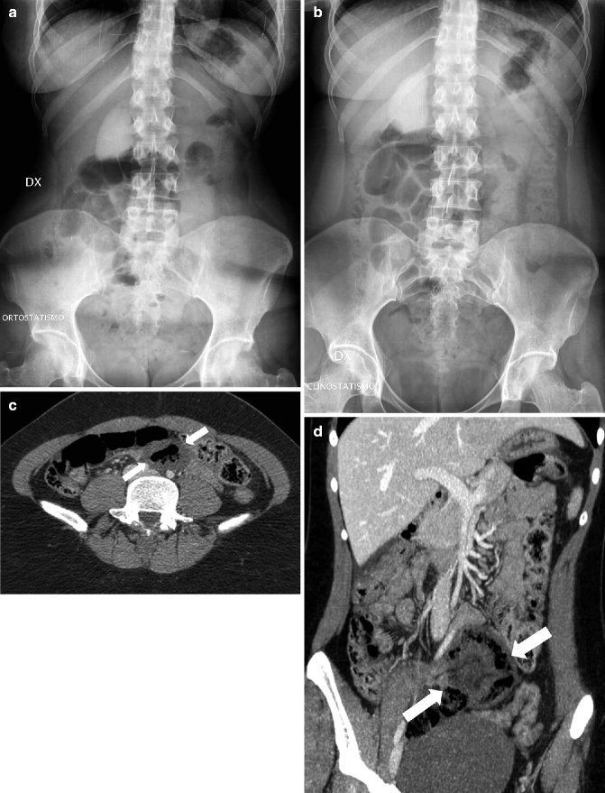
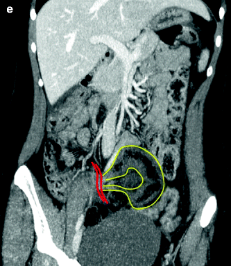
Fig. 29.4
(a–e) Closed-loop obstruction. Discrepancy between the radiographic picture and CT findings in a patient with untreatable acute abdominal pain. Upright (a) and anteroposterior (b) radiographs show overdistension of ileal loops in the right and middle quadrants without thickened bowel walls and/or air-fluid levels. (c) Axial contrast-enhanced MDCT scan shows dilated ileal loop with marked decreased enhancement of the bowel wall (arrows). (d) Maximum intensity projection image demonstrates a closed loop in the middle lower abdominal quadrant (arrows). (e) Same CT scan as (d), with yellow lines demonstrating the closed loop and red lines indicating the “beak” of the closed loop at the site of obstruction. At surgery, an adhesional band was found and the infarcted ileal loop was resected
29.1.1.8 Intussusception
It is an unusual cause of bowel obstruction consisting of an invagination of a proximal segment of bowel (intussusceptum) into the lumen of a distal, adjacent segment of bowel (intussuscipiens). Intussusception may involve only the small bowel (ileoileal intussusception, typically in adults), the small and the large bowel (ileocolic intussusception, typically in children), or just the colon (colocolic intussusception). In adults, intussusception must be considered as a complication of a neoplasm, until proven otherwise. On the basis of canalization, three types of intussusception are identifiable: (1) cold intussusception (asymptomatic), (2) incomplete and reversible hot intussusception (with recurrent and transient episodes of intestinal obstruction), and (3) complete and irreversible hot intussusception (complicated obstruction due to simultaneous involvement of both the bowel and its vascular supply) (Fig. 29.5).
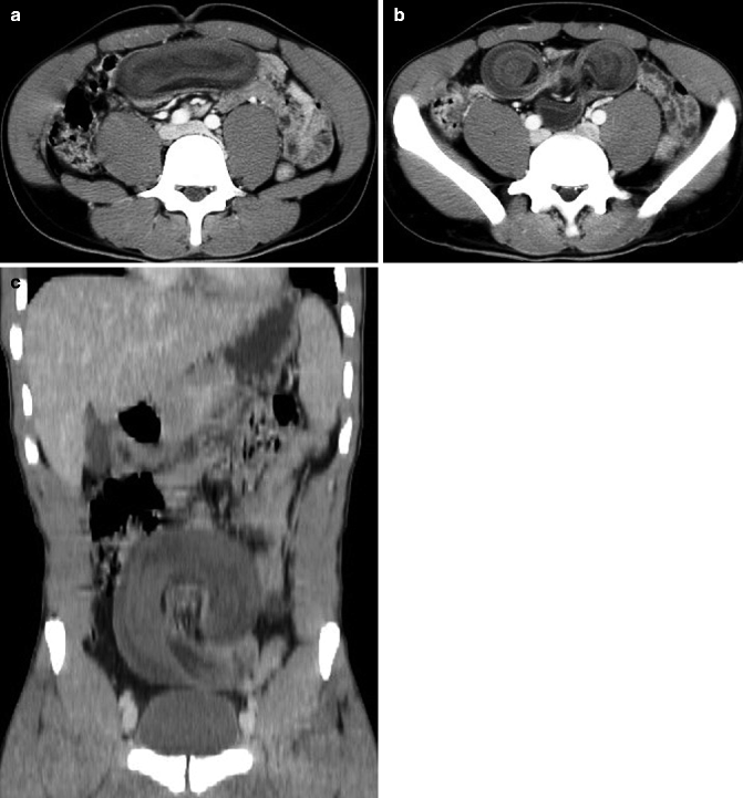
Fig. 29.5
(a–c) Hot intussusception. Ileoileal intussusception in both the longitudinal (a) and axial axes (b) with typical “target” appearance (b) and markedly reduced bowel wall enhancement, causing small bowel obstruction complicated by ischemia. (c) Maximum intensity projection image demonstrates ileal volvulus in the middle abdominal quadrants, complicating the hot intussusception
29.1.2 Imaging
29.1.2.1 Radiographic and Ultrasound Findings of Simple SBO
Dilated proximal small bowel loops
Multiple air-fluid levels
Collapsed distal loops
Absence of peritoneal fluid (US)
29.1.2.2 CT Findings of Simple SBO
Dilated loops proximally to the transition zone.
Transition zone (point of obstruction) is the abrupt caliber change between the dilated and the collapsed loops.
Collapsed loops distally to the point of obstruction.Related posts:

Stay updated, free articles. Join our Telegram channel

Full access? Get Clinical Tree