High-quality images are critical for accurate image interpretation. The first article by Drs DeMartini and Rahbar reviews technical considerations for performing high-quality breast MRI at both 1.5 T and 3 T, recognizing that clinical breast MRI at 3 T is increasing. This article concludes with a discussion of the American College of Radiology Breast MR Accreditation Program, which opened in 2010. Article two by Drs Edwards, Lipson, Ikeda, and Lee is devoted to the BI-RADS lexicon, which has helped to improve consistency in breast MR interpretation and clarity of reporting. Expected updates and revisions to the lexicon, such as the reporting of background parenchymal enhancement, are included.
The next 5 articles cover clinical indications for breast MRI. Breast MRI is currently considered the most sensitive imaging tool for breast cancer detection. High-risk screening is one of the accepted clinical indications for breast MRI and represents a growing percentage of the breast MRI volume in clinical practices. Article 3 by Drs Mahoney and Newell presents a comparison of breast MRI vs breast ultrasound for screening high-risk women, while article 4 focuses on the risk factors that would place a woman into the high-risk category. Article 4 by Drs Dershaw and Sung also reviews recent literature suggesting benefit of breast MRI screening for subgroups of women at intermediate risk, such as those with a personal history of breast cancer or lobular carcinoma in situ. Articles 5 and 6 move away from screening and focus on the evaluation of patients with known breast cancer. Extent of disease evaluation was one of the early applications for breast MRI but is currently the most controversial. Specific scenarios in which MRI may be beneficial in the newly diagnosed cancer patient are reviewed in Article 5 by Drs Brasic, Wisner, and Joe. On the other hand, breast MRI is considered superior to mammography or ultrasound for monitoring response of locally advanced breast cancer to neoadjuvant chemotherapy and the type of treatment response seen at MRI may be predictive of overall and disease-free survival as detailed in Article 6 by Drs Ojeda-Fournier, de Guzman, and Hylton. Moreover, breast MRI is useful for presurgical planning to assess patient’s candidacy for breast conservation at the end of neoadjuvant treatment.
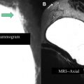
Article 7 by Dr Brenner covers a noncancer application of breast MRI, namely evaluation of the integrity of silicone implants. While saline implant rupture is usually clinically apparent, silicone implant rupture evaluation requires dedicated MRI protocols specific for silicone signal and which include imaging in orthogonal planes.
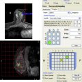
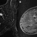
The last part of this issue is devoted to advanced topics in breast MRI. Article 8 by Drs Comstock and Sung provides a thoughtful discussion of the use of BI-RADS 3 “Probably Benign” assessment at breast MRI. This is a challenging topic given the paucity of data for guidance. Classically benign findings, which should be classified as BI-RADS 2 rather than BI-RADS 3, are also presented. Article 9 by Dr Price covers breast MRI biopsy step by step, including tips and tricks for difficult or complex cases. As with any other image-guided biopsy, it is important to assess for radiologic-pathologic concordance. Because there is no “real-time” visualization and no specimen radiograph for MRI-guided biopsy, radiologic-pathologic correlation can be more challenging. Article 10 by Drs Heller, Hernandez, and Moy provides a review of radiologic-pathologic correlation at breast MRI with a focus on the appropriate management for high-risk lesions. Articles 11 and 12 end this issue with a discussion of emerging breast MRI techniques, sometimes referred to as “functional MRI.” Article 11 by Drs Partridge and McDonald covers the topic of diffusion-weighted imaging, a noncontrast technique with potential to improve specificity of breast MRI by characterizing a lesion as more likely benign or malignant based on its diffusion characteristics. Article 12 by Dr Bolan reviews the current status of and potential applications for MR spectroscopy, a noncontrast technique for measuring metabolic biomarkers of malignancy such as choline.
I would like to thank all the contributors from the breast MRI community who generously shared their time and expertise to write informative and clinically relevant articles. Thank you to Pamela Hetherington and the rest of the staff at Elsevier, who helped to keep us all organized and on time with this publication. And, of course, a big thank you to my family for their support and indulgence when I needed “quiet time” for this project.
Stay updated, free articles. Join our Telegram channel

Full access? Get Clinical Tree


