and Vincent L. Sorrell2
(1)
Division of Nuclear Medicine and Molecular Imaging Department of Radiology, University of Kentucky, Lexington, Kentucky, USA
(2)
Division of Cardiovascular Medicine Department of Internal Medicine Gill Heart Institute, University of Kentucky, Lexington, Kentucky, USA
Electronic supplementary material
The online version of this chapter (doi: 10.1007/978-3-319-25436-4_6) contains supplementary material, which is available to authorized users.
Review of the raw projection data from routine MPI may reveal incidental breast pathology (Table 6.1) (Gedik et al. 2007; Raza et al. 2005a, b). The left and most of the right breast are included in the field-of-view during routine SPECT MPI. A variety of benign conditions tend to show a diffuse and symmetric pattern (Fig. 6.1) (Chamarthy and Travin 2010). Lactating breasts (Fig. 6.2) will show varying degrees of bilaterally symmetric increased uptake depending on the postpartum state and breastfeeding status (Ramakrishna and Miller 2004). Breastfeeding does not need to be stopped for the examination; 99mTc radiopharmaceuticals are preferred in this setting (Hesse et al. 2005). In males, gynecomastia may be evident (Fig. 6.3) and is often associated with underlying liver disease or associated with hormonal therapy or medical therapy for congestive heart failure (e.g., spironolactone).


Table 6.1
Differential diagnosis of “hot” and “cold” imaging findings related to the breasts
Organ system | “Hot” finding | “Cold” finding | References |
|---|---|---|---|
Breasts | Cystic disease Fibroadenoma Microcalcifications Hematoma Scar Lactation Gynecomastia (periareolar pattern) Carcinoma | Cyst Implants Prosthesis Soft-tissue/shifting breasts (positional) Mastectomy | Arbab et al. (1995) Burrell and MacDonald (2006) Chamarthy and Travin (2010) García-Talavera et al. (2013) Gedik et al. (2007) Hendel et al. (1999) Hesse et al. (2005) Holly et al. (2010) Howarth et al. (1996) Mlikotic and Mishkin (2000) Ramakrishna and Miller (2004) Shih et al. (2002) Waxman (1997) Williams et al. (2003) |

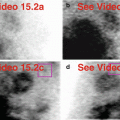
Fig. 6.1
Symmetric, diffusely “hot” breasts. The breast uptake is relatively intense and extensive but localizes to the central subareolar tissues (a–d). A 47-year-old female has benign-appearing “dense” breasts by ultrasound; she opted out of conventional mammography.(a) Stress raw projection images (Video 6.1a, frame 1), 99mTc sestamibi. (b) Stress raw projection image (Video 6.1b, frame 1, RAO), 99mTc sestamibi, both breasts (yellow ovals). (c) Stress raw projection image (Video 6.1b, frame 25, Anterior), 99mTc sestamibi, left breast contour (yellow lines) extends well beyond internal breast uptake. (d) Stress raw projection image (Video 6.1b, frame 44, LAO), 99mTc sestamibi, both breasts (right, yellow oval; left, pink oval)

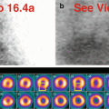
Fig. 6.2
Lactation. Diffusely “hot” breasts (a, b) in a lactating 38-year-old female. The anterolateral extracardiac activity has much greater relative intensity on rest processed images (c); this pattern is likely related to relative differences in cardiac-to-breast activity after treadmill exercise. (a) Rest raw projection images (Video 6.2a, frame 1), 99mTc tetrofosmin; breasts appear spread out due to arms raised and supine position for imaging. (b) Rest raw projection image (Video 6.2b, frame 18, Anterior), 99mTc tetrofosmin, breasts (yellow ovals); note left breast overlies left ventricle. (c) Stress/rest processed SPECT images (SA, HLA, VLA) (without and with AC), left breast uptake (yellow boxes on representative images)

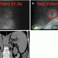
Fig. 6.3
Gynecomastia. There is well-defined localized periareolar activity creating “hot” breasts (a–c) in a cirrhotic male. Note the “hot” stomach which is related to the associated gastropathy (b). The left breast activity is anteriorly situated relative to the heart (c).(a) Stress raw projection images (Video 6.3a, frame 1), 99mTc sestamibi. (b) Stress raw projection image (Video 6.3b, frame 18, Anterior), 99mTc sestamibi, areolar regions of both breasts (blue ovals), stomach (pink lines). (c) Stress raw projection image (Video 6.3b, frame 62, Lateral), 99mTc sestamibi, areolar region of left breast (blue circle), left ventricular myocardium (yellow lines)
99mTc sestamibi has been long used clinically to evaluate the breast for malignancy (Taillefer et al. 1995, 1998; Waxman 1997). Breast-specific gamma imaging (BSGI) remains under active investigation as a molecular approach adjunctive to mammography, ultrasonography, and magnetic resonance imaging, particularly in mammographically dense breasts (Holbrook and Newel 2015). Thus, the differential diagnosis for focal increased uptake in the breasts includes primary, metastatic, or recurrent breast malignancy, particularly if it is unilateral and intensely avid for 201Tl chloride and 99mTc sestamibi (Fig. 6.4) (Arbab et al. 1995; Chamarthy and Travin 2010; García-Talavera et al. 2013; Hendel et al. 1999; Mlikotic and Mishkin 2000; Shih et al. 2002; Williams et al. 2003). The rest raw acquisition might be more revealing and should be reviewed, ideally with a whole-field-of-view reconstruction for three-dimensional characterization of a suspected lesion (Seo et al. 2005). The written report should include wording along the lines of:


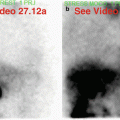
Fig. 6.4


Right breast cancer. A 64-year-old female has a diffusely “hot” right breast. (a) Stress raw projection image, 99mTc sestamibi, right breast (yellow box), heart (blue circle)
Stay updated, free articles. Join our Telegram channel

Full access? Get Clinical Tree


