Bronchial Atresia
Jud W. Gurney, MD, FACR
Key Facts
Terminology
Congenital atresia of segmental bronchus with normal distal architecture
Imaging Findings
Apicoposterior segment left upper lobe (50%), followed by right upper lobe (20%), lower lobes (15% each), and rarely right middle lobe (< 5%)
Typical triad
Central nodule or mass representing mucoid impaction distal to atretic bronchus (bronchocele)
Hyperlucency of affected segment
Hypoperfusion of affected segment with paucity of vessels
Bronchocele
Blunt horn-like protrusions distal to mass (mucoid impaction in bronchiectatic bronchi)
Occasionally have air-fluid level
Top Differential Diagnoses
Mucoid Impaction with Hyperinflation
Congenital Lobar Emphysema
Intralobar Pulmonary Sequestration
Pathology
2nd most common congenital tracheobronchial malformation after pulmonary sequestration
2 theories of pathogenesis
Disconnected cells from bronchial bud
Intrauterine vascular injury
Diagnostic Checklist
Exclude slow-growing endobronchial tumor
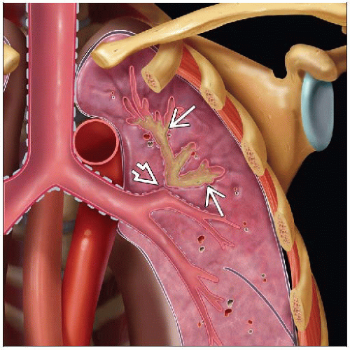 Coronal graphic shows bronchial atresia  with branching bronchocele with branching bronchocele  in the apical segment left upper lobe. in the apical segment left upper lobe. |
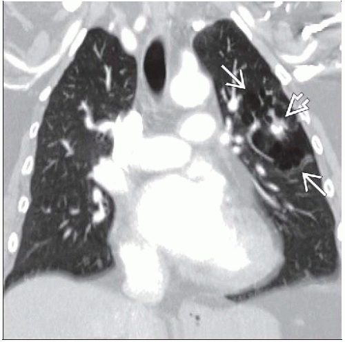 Coronal CECT shows focal lucency  from bronchial atresia and central mucus-filled bronchocele from bronchial atresia and central mucus-filled bronchocele  in this patient with segmental bronchial atresia. in this patient with segmental bronchial atresia. |
TERMINOLOGY
Abbreviations and Synonyms
Mucocele
Definitions
Congenital atresia of segmental bronchus with normal distal architecture
IMAGING FINDINGS
General Features
Best diagnostic clue: Round, sharply-defined, perihilar mass with distal hyperinflation
Patient position/location
Apicoposterior segment left upper lobe (50%)
Followed by right upper lobe (20%), lower lobes (15% each) and rarely right middle lobe (< 5%)
Size: Bronchocele usually > 1 cm in diameter
Morphology: Atretic bronchus usually segmental but may be lobar or distally in subsegmental airways
CT Findings
Triad: Bronchocele and hyperlucent and hypoperfused segment diagnostic of bronchial atresia
Bronchocele
May be of lower attenuation due to mucoid material
May contain calcium
No enhancement
Shape: Tubular, spherical, branching
Bronchocele aligned along central axis of affected segment
Bronchocele located medial to hyperinflated lung
Wedge-shaped hyperinflated lung surrounds bronchocele
Rare systemic arterial supply (bronchoarterial malinosculation)
Radiographic Findings
Radiography
Typical triad
Central nodule or mass representing mucoid impaction distal to atretic bronchus (bronchocele)
Hyperlucency of affected segment
Hypoperfusion of affected segment with paucity of vessels
Bronchocele
Sharply defined rounded or tubular branching opacities adjacent to the hilum (teardrops, grape-like clusters, gloved-finger appearance)
Bronchocele points towards hilum
Blunt horn-like protrusions distal to mass (mucoid impaction in bronchiectatic bronchi)
Occasionally have air-fluid level
Hyperlucent lobe
Neonates: Lobe or segment distal to atretic bronchus fluid-filled, gradually replaced by air
Resorption of fluid occurs within 1st week of life
Ventilation from collateral air drift via intraalveolar pores of Kohn and bronchoalveolar channels of Lambert across incomplete intrapulmonary fissures
Boomerang sign
Parabolic curve: Junction of hyperinflated segment with adjacent normal lung
Associated findings
Hypoplastic ribs, pectus excavatum
Ultrasonographic Findings
Can be detected in utero
Fluid-filled upper lobe
Differential
Cystic adenomatoid malformation
Congenital diaphragmatic hernia
Bronchopulmonary foregut malformations
Congenital lobar emphysema
Nuclear Medicine Findings
V/Q scan
Hypoperfusion of affected segment
Delayed ventilation of affected segment with delayed washout (air-trapping)
Imaging Recommendations
Best imaging tool: CT procedure of choice to characterize bronchocele, airway anatomy, and distal hyperinflated lung and other associated anomalies
Protocol advice
Expiratory CT demonstrates accentuated hyperinflation of affected segments
Multidetector CT useful in demonstrating anatomy of atretic bronchus
DIFFERENTIAL DIAGNOSIS
Mucoid Impaction with Hyperinflation
Bronchial atresia
Intralobar sequestration
Intrapulmonary bronchogenic cyst
Congenital Lobar Emphysema
No bronchocele
Left upper lobe also most commonly affected
Hyperinflated lobe causes mass effect with shift of mediastinum away from affected lobe
Usually diagnosed in infancy with respiratory distress
Intralobar Pulmonary Sequestration
May have distal hyperinflation
Abnormal systemic arterial supply, usually from aorta
Most common location is left lower lobe in paravertebral angle
Intrapulmonary Bronchogenic Cyst
Usually located in medial 1/3 of lung in lower lobes
May have distal hyperinflation
Cyst may be fluid-filled, air-filled, or both (air-fluid level)
Arteriovenous Malformation
Abnormal feeding artery and draining vein
Nodule will enhance with contrast administration
No bronchial obstruction, no hyperlucency or hyperinflation
Mucoid Impaction Associated Conditions
Allergic Bronchopulmonary Aspergillosis
Central bronchiectasis with mucoid impaction
Diffuse, not localized
Primarily affects upper lung zones
Distal lung usually abnormal
Small airways disease: Tubular branching opacities, hyperinflation
Cystic Fibrosis
Central bronchiectasis
Bilateral disease usually more severe in upper lung zones, especially right upper lobe
May have mucoid impaction
Distal lung usually abnormalRelated posts:

Stay updated, free articles. Join our Telegram channel

Full access? Get Clinical Tree


