Fig. 27.1
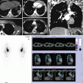
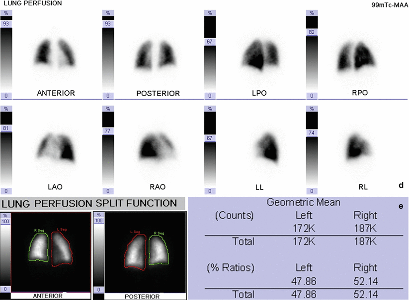
(a–c) Chest X-ray (a) and coronal (b) and axial (c) CT scan of the chest show normal findings, the absence of parenchymal or pleural abnormalities, regular airway. (d–e) Lung perfusion scan: normal tracer uptake and normal distribution are evident in both lungs showing symmetrical function. Lung perfusion scan ruled out pulmonary embolism
27.4.2 Case 27.2: Lung Perfusion and Ventilation Scan – Normal Pattern in Newborn
A 9-month-old infant presenting persistent wheezing since the first month of life, treated by asthma therapy and leukotriene. Negative sweat test on two occasions. At 9 months of life, a worsening dyspnea and bronchospasm episode occurred, and chest X-ray showed atelectatic areas with air trapping and bilateral consolidations, especially in the right hemithorax.
A further examination of the chest by CT angiography showed tracheal bronchus (upper right of lobar bronchus that departs from the right edge of the lower third of the trachea), while lung perfusion and ventilation scan revealed a normal perfusion and pulmonary ventilation. Respiratory physiotherapy has been associated to medical therapy, and the child no longer had episodes of bronchospasm (Fig. 27.2).
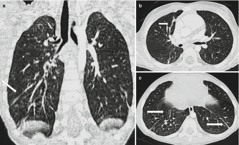
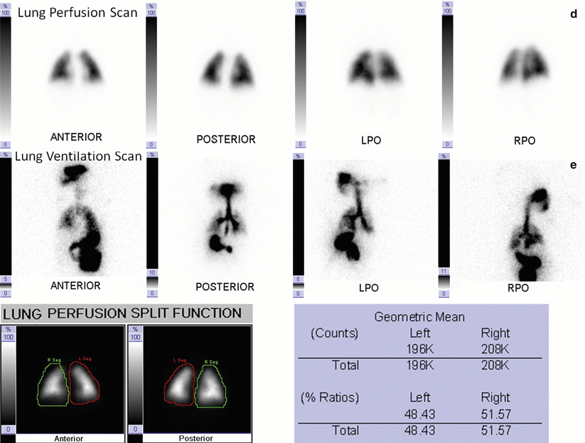


Fig. 27.2
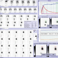
(a–c) CT coronal view image of the chest (a) shows upper right lobar bronchus that departs from the right edge of the lower third of the trachea (tracheal bronchus, black arrow). Images in the axial view (b) show streaks of laminar atelectasis in the front segments of the right upper lobe and apical right lower lobe and in the basal segments of the posterior side of the lower lobes bilaterally (c) (white arrows). (d–e) In the first row (d) is evident normal tracer uptake and distribution in both lungs showing symmetrical function. In newborn, lung perfusion scan can be performed by only four views (anterior, posterior, left, and right posterior obliques). In the second row (e), normal distribution of ventilation is evident in both lungs. To perform a good ventilation scan in newborn, an adequate mask is necessary eliciting infant crying
27.4.3 Case 27.3: Lung Ventilation Scan for Measuring Mucociliary Clearance – Normal Pattern
An 8-year-old girl with frequent respiratory infection. Chest X-ray was performed showing diffuse thickening of the bronchial walls bilaterally with lung hyperinflation in the absence of areas of parenchymal consolidation. In suspicion of primary ciliary dyskinesia, the child underwent nasal brushing which showed a nondiagnostic result. Lung ventilation scan for measuring mucociliary clearance was required, revealing a normal pattern (Fig. 27.3).
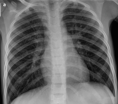
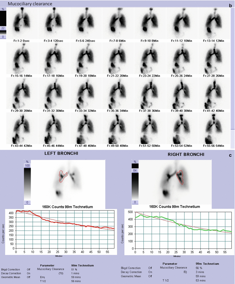


Fig. 27.3
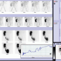
(a). Chest X-ray image in the front projection shows diffuse thickening of the bronchial walls bilaterally with lung hyperinflation in the absence of areas of parenchymal consolidation. (b) By visual assessment, normal distribution of ventilation is evident in both lungs. Regular particle deposition and normal mucociliary clearance from the respiratory airways are evident in both lungs. (c) A/T curve analysis shows a bilaterally rapid decrement of activity, resulting in normal quantitative clearance parameters in 60 min
27.4.4 Case 27.4
A 9-day-old newborn with good general condition at birth and cardiorespiratory parameters within normal limits. At clinical skill, reduced air penetration at the upper and middle fields of the right lung. He was submitted to chest X-rays at first and then to chest-CT and lung perfusion scan for better definition of the clinical condition and therapy (Fig. 27.4).
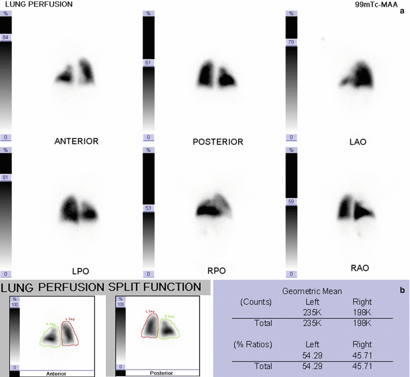

Fig. 27.4
(a–b) Normal left lung perfusion. Hypoperfusion in the entire upper and middle lobe of the right lung due to the hypoventilation and regular perfusion of the lower lobe. (c–f). Chest X-ray front view (c) displays extensive hyperlucency area in right lung, with thinning bronchovascular markings (black arrow) and bundling in lung plot in the baseline paracardiac (asterisk). Chest CT image in coronal reconstructions MIP (d) shows right upper lobar bronchus originating from the distal third of the trachea (tracheal bronchus, white arrow) with hypodense parenchymal lung’s right upper lobe due to air trapping, as also shown in the TC axial (e) and coronal (f) images (black arrows)
27.4.5 Case 27.5: Lung Embolism in Child Affected by LES
A 13-year-old girl affected by SLE treated by steroid therapy. She had more hospitalizations for pain in the anterior chest region during which echocardiographic controls resulted normal. After several months, she showed left lower limb thrombophlebitis associated with dyspnea. During hospitalization, in suspicion of lung thromboembolism, a chest CT and a lung perfusion scan were performed. He was hospitalized once again in spite of crisis characterized by “air hunger,” which was interpreted as panic attack (chest CT and lung scan performed during hospitalization were normal) (Fig. 27.5).
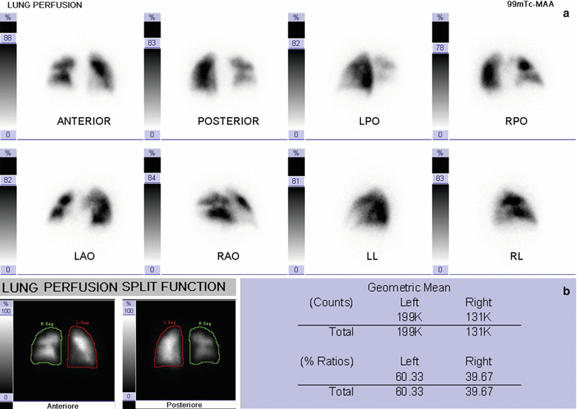
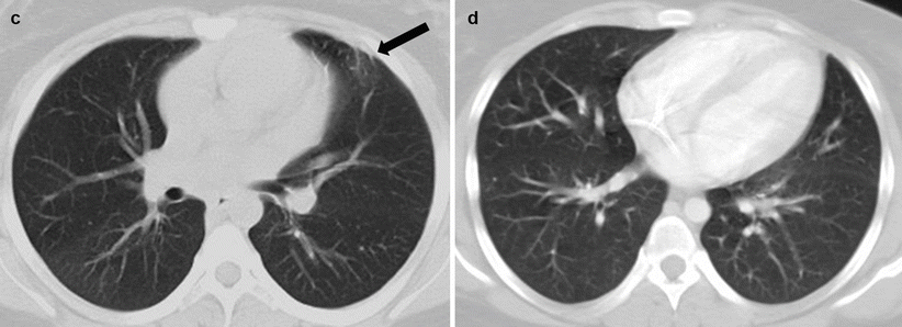
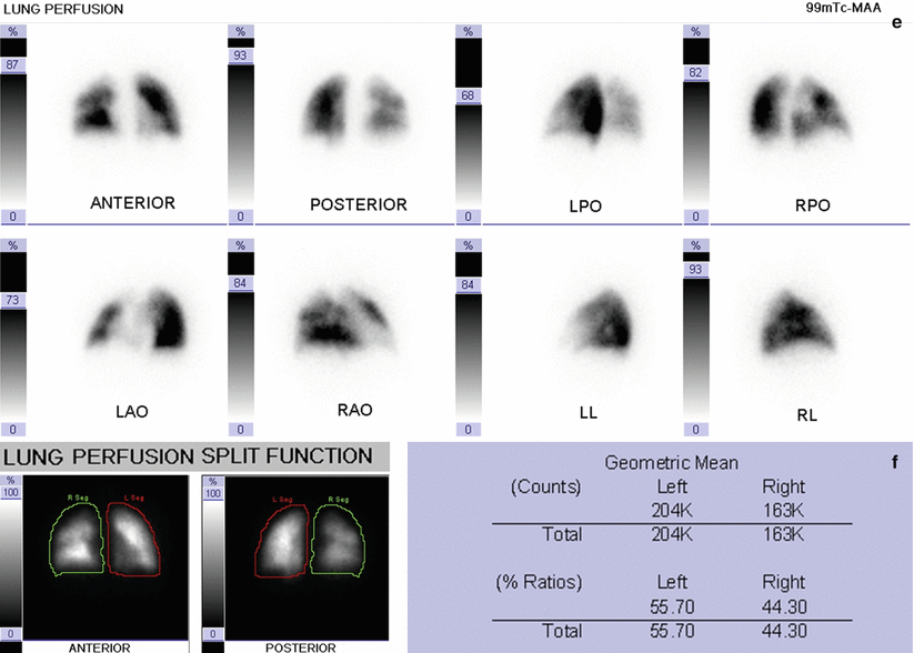


Fig. 27.5
(a–b) Lung perfusion scan in the course of embolism showed multiple segmental perfusion deficits in the right lung which showed associated reduced uptake (quantitative perfusion parameters: 60.33 % left lung and 39.67 % right lung). Axial CT image (c) shows mild heterogeneity of parenchymal density with relative hyperdensity of the apical segment of the right lower lobe and striations dense front subppleuriche left fibro-disventilative (black arrow). Axial CT image (d) of distant examination performed 2 years later shows substantial stability of the finds with increasingly evident parenchymal inhomogeneous density and areas of low attenuation lobular air trapping. (e–f) Lung perfusion scan showed increased perfusion in the right lung with better distribution of the radiopharmaceutical uptake (quantitative perfusion parameters: 55.70 % left lung and 44.30 % right lung)
27.4.6 Case 27.6: A Case of Acute Respiratory Distress at Birth
A 4-month-old baby who presented at birth cyanosis and cardiorespiratory depression. She needed resuscitation in the delivery room. The diagnosis was hyaline membrane disease, tracheomalacia, and suspected ciliary dyskinesia. At 2 months of life, she was intubated for 45 days. O2 therapy has been required.
The child reached our hospital for a sudden worsening of respiratory symptoms, and she was admitted into the intensive care unit. The patient was placed on mechanical ventilation. During mechanical ventilation, sudden worsening of the general conditions with dyspnea and cyanosis crisis occurred. She was submitted to chest CT and lung perfusion scan for clarifying the pulmonary framework (Fig. 27.6).
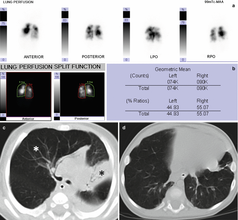

Fig. 27.6


(a–b) Lung perfusion scan shows multiple areas of reduced or absent perfusion signal in both lungs, extending from the apical zones to the pulmonary bases; only few defects had segmental shape. The quantitative evaluation of pulmonary perfusion is symmetrical as follows: Left lung = 44.9 %; Right lung Dx = 55.1 %. This scintigraphic framework suggested widespread disease of lung parenchyma as often observed in patients with bronchodysplasia. (c–d) Axial CT images of the thorax (c) show widespread disease of lung parenchyma with hypodiafania due to hyperinflation of the middle lobe (white asterisk), partial atelectasis of left upper lobe (black asterisk), and of right upper lobe with mediastinal shift to the left, and evidence of striae, consolidations, and parenchymal distortion (d)
Stay updated, free articles. Join our Telegram channel

Full access? Get Clinical Tree


