Presentation and Presenting Images
( ▶ Fig. 49.1, ▶ Fig. 49.2, ▶ Fig. 49.3, ▶ Fig. 49.4)
A 81-year-old female presents for screening mammography. She has a personal history of treated bilateral breast cancers. Her most recent breast cancer (ductal carcinoma in situ [DCIS]) was 2 years ago involving the left breast, and was treated with a lumpectomy and radiation therapy.
49.2 Key Images
( ▶ Fig. 49.5, ▶ Fig. 49.6, ▶ Fig. 49.7)
49.2.1 Breast Tissue Density
There are scattered areas of fibroglandular density.
49.2.2 Imaging Findings
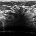
There is subareolar architectural distortion and retraction, which is marked with a skin line marker that denotes the prior lumpectomy scar. Adjacent to the region, there are grouped linear calcifications ( ▶ Fig. 49.6 and ▶ Fig. 49.7). In addition, there is a possible focal asymmetry at the 1 o’clock location in the middle depth ( ▶ Fig. 49.5 and ▶ Fig. 49.6).
49.3 BI-RADS Classification and Action
Category 0: Mammography: Incomplete. Need additional imaging evaluation and/or prior mammograms for comparison.
49.4 Diagnostic Images
( ▶ Fig. 49.8, ▶ Fig. 49.9, ▶ Fig. 49.10, ▶ Fig. 49.11, ▶ Fig. 49.12, ▶ Fig. 49.13)
49.4.1 Imaging Findings
The diagnostic imaging resolved the asymmetry ( ▶ Fig. 49.8 and ▶ Fig. 49.9) in the upper outer quadrant which when compared to prior mammograms appeared to represent a summation artifact. The magnification images further define the 1-cm area of calcifications located at 4 o’clock in the middle depth ( ▶ Fig. 49.10, ▶ Fig. 49.11, and ▶ Fig. 49.12). These calcifications are fine-linear branching calcifications and are located 1.7 cm inferior to the center of the lumpectomy bed. The other scattered calcifications are stable. A biopsy of these calcifications was performed with calcifications found in several cores ( ▶ Fig. 49.13).
49.5 BI-RADS Classification and Action
Category 4C: High suspicion for malignancy
49.6 Differential Diagnosis
Summation artifact and DCIS: The possible asymmetry appeared stable and less worrisome on additional imaging and further comparison to prior imaging studies. The calcifications are in a linear distribution and are suspicious. The biopsy yielded high grade DCIS, which is similar to her prior cancer in this breast.
Fat necrosis: The calcifications of fat necrosis can mimic those of carcinoma. Typically fat necrosis presents as dystrophic calcifications. These calcifications are suspicious and warrant a biopsy.
Secretory calcifications: There are no other secretory calcifications in the breast. These calcifications are too fine and branching to be dismissed as benign.
49.7 Essential Facts
The calcifications in this case would have been detected with or without the digital breast tomosynthesis (DBT) evaluation.
The detection of microcalcifications on DBT is affected by the detector type, the image acquisition and reconstruction parameters, and blur due to source, detector, or patient motion during acquisition.
The detection of invasive cancers is increased with the implementation of DBT. The retrospective multi-site study by Friedewald and colleagues (2014) observed a 41% relative increase in the cancer detection rate from 2.9 to 4.1 per 1000. In this same study the detection of DCIS remained unchanged at 1.4 per 1000.
Early investigation suggests that the synthetic mammogram (reconstructed from the DBT images) performs equally well when compared to the full-field digital mammogram (FFDM). The benefit is in a reduction in patient radiation dose due to performing only DBT and not FFDM and DBT.
49.8 Management and Digital Breast Tomosynthesis Principles
DBT when included in mammographic screening may add value by increasing the cancer detection rate for invasive cancers, optimizing patient outcomes, .
DBT is well suited to the assessment of asymmetries. Most asymmetry recalls are summation artifacts, which would be effaced by DBT.
Due to inherent technical factors DBT is less sensitive for the detection of calcifications compared to FFDM.
49.9 Further Reading
[1] Das M, Gifford HC, O’Connor JM, Glick SJ. Evaluation of a variable dose acquisition technique for microcalcification and mass detection in digital breast tomosynthesis. Med Phys. 2009; 36(6): 1976‐1984 PubMed
[2] Friedewald SM, Rafferty EA, Rose SL, et al. Breast cancer screening using tomosynthesis in combination with digital mammography. JAMA. 2014; 311(24): 2499‐2507 PubMed
[3] Zuley ML, Guo B, Catullo VJ, et al. Comparison of two-dimensional synthesized mammograms versus original digital mammograms alone and in combination with tomosynthesis images. Radiology. 2014; 271(3): 664‐671 PubMed

Fig. 49.1 Left craniocaudal (LCC) mammogram.
Stay updated, free articles. Join our Telegram channel

Full access? Get Clinical Tree


