Carcinoma of Unknown Primary: Including Paraneoplastic Neurological Syndromes
Jennifer Rodriguez-Ferrer
Richard L. Wahl
Carcinoma of unknown primary (CUP) accounts for 3% to 5% of all new cancer diagnoses. It is the seventh most common malignancy and the fourth most common cause of cancer death in both males and females. The median age at presentation is 60, with CUP more frequent in men than in women. The prognosis of patients with CUP is generally poor. The median survival rate is approximately 3 to 11 months with less than 25% of patients surviving over 1 year. Survival rates differ among histological subgroups and are worse in those patients who present with widely disseminated disease (1,2,3).
CUP is defined as histologically confirmed metastatic cancer with no identifiable primary site after a complete history and physical examination (including a pelvic and rectal examination), complete blood count, biochemistry, urinalysis, occult fecal blood test, serum tumor markers, histopathology review of biopsy material with the use of immunohistochemistry, mammography in females, computed tomography (CT) of the chest, abdomen and pelvis, or any other relevant test. Until recently, 18-F-fluoro-2-deoxy-D-glucose (FDG) positron emission tomography (PET) was not one of these tests. There is some variability in the literature as to the intensity of the work-up for CUP, likely contributing to some variability in the rates of identification of the previously occult primary lesions by imaging tests like PET. In the majority of cases the primary site will never be found during the patient’s lifetime using a work-up that does not include PET. Twenty percent of patients have no detectable primary tumor site even at postmortem studies. Conventional radiological imaging identifies about 20% to 27% of the primary tumors after intensive investigation. Most commonly, patients seek medical attention because of the recent enlargement of a superficial lymph node or present with visceral masses, bone lesions either symptomatic or detected at radiographic examinations for an unrelated indication. Possible explanations for the inability to detect the primary tumor include: (a) spontaneous regression (the primary is not detectable when metastasis becomes evident, not a common phenomenon but this process has been described in several tumors such as melanoma; approximately 5% of patients with malignant melanoma present without a identifiable primary site); (b) immune- modulated destruction of the primary cancer; or (c) the primary tumor’s malignant phenotype and genotype favor metastatic ability over local tumor growth (1,2,3).
CUPs are categorized into four major subtypes by routine light microscopy criteria:
(a) adenocarcinomas, well to moderately differentiated 50% to 60%; (b) poorly differentiated adenocarcinomas or carcinomas 30% to 40%; (c) squamous cell carcinoma (SCC) 5% to 8%; and (d) undifferentiated neoplasms 2% to 5% (including lymphoma, sarcoma, melanoma, primitive neuroendocrine tumors, germ cell tumors, and embryonal tumors). Patients with poorly differentiated carcinomas and metastatic adenocarcinoma of unknown primary have the poorest prognosis, while metastatic SCC has a more favorable prognosis. CUP has an unpredictable metastatic pattern that differs from the pattern distribution seen in tumors with identifiable primary lesions. For example, patients eventually diagnosed with prostate cancer who present as CUP have a high incidence of metastases to nonosseous sites such as lung, liver, and brain. Bone metastases are less common than lung metastases in thyroid cancer presenting as CUP than in other presentations of thyroid cancer.
CUP falls into two major categories of anatomical presentation: those with metastatic involvement to lymph nodes or patients presenting with visceral tumor involvement. As discussed later, CUP can also present with neurological syndromes. The most common sites for involvement of metastatic disease are lymph nodes, liver, lung, or bone. Lymph nodes in the cervical and supraclavicular region are the most common metastatic sites for CUP. Histologically, these tumors are usually SCC, but occasionally may be adenocarcinoma, melanoma, or anaplastic tumors. If the metastatic lymph nodes are located in the upper and middle cervical levels, especially in the case of SCC, a primary tumor of the head and neck region is highly suspected. If the lower cervical lymph nodes are only involved, the primary tumor is often located below the clavicles. Also, cervical metastases of non-SCC at any level can be due to a primary tumor located outside the head and neck area, and most
likely the primary is found in the lung, gastrointestinal, or urogenital system. Although axillary nodal involvement in a middle-aged women is highly suggestive of primary breast carcinoma, nonpalpable breast cancer presenting as axillary metastases is quite rare, accounting for less than 0.5% of all breast cancers (although since breast cancers are quite common, this presentation of CUP is not infrequent). Metastases to inguinal nodes more commonly originate from primary tumors of the anogenital area, where diffuse metastatic involvement of the peritoneum or retroperitoneal lymph nodes are most consistent with primary tumors of the ovary, peritoneum, prostate, or testis. Pulmonary and hepatic metastases may arise from any site, whereas the most common origin of bone metastases are thyroid, breast, and prostate cancers (1,2,3).
likely the primary is found in the lung, gastrointestinal, or urogenital system. Although axillary nodal involvement in a middle-aged women is highly suggestive of primary breast carcinoma, nonpalpable breast cancer presenting as axillary metastases is quite rare, accounting for less than 0.5% of all breast cancers (although since breast cancers are quite common, this presentation of CUP is not infrequent). Metastases to inguinal nodes more commonly originate from primary tumors of the anogenital area, where diffuse metastatic involvement of the peritoneum or retroperitoneal lymph nodes are most consistent with primary tumors of the ovary, peritoneum, prostate, or testis. Pulmonary and hepatic metastases may arise from any site, whereas the most common origin of bone metastases are thyroid, breast, and prostate cancers (1,2,3).
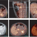
FDG PET has been used for the evaluation of patients with CUP, particularly in patients with isolated neck metastases, but very few data are available on its usefulness in extracervical metastases. The detection rates in the literature for FDG PET have ranged widely, from 8% to 57% (Table 8.22.1). This wide range is attributed to the different definitions used for CUP between studies, differing make up of the patient populations, differing techniques (e.g., PET vs. PET/CT), and likely differing reader thresholds for positivity. Overall, the PET detection rate of primary lesions is about 39% (4,5,6,7,8,9,10,11,12,13,14,15,16,17,18,19,20).
Related posts:
Stay updated, free articles. Join our Telegram channel

Full access? Get Clinical Tree


