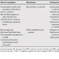
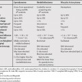
66 A false aneurysm of the left ventricle is a rare complication of myocardial infarction. A false aneurysm is a contained rupture; the wall does not contain myocardial elements. It is important to differentiate a false aneurysm from a true aneurysm, which has a wall containing myocardial elements, as a false aneurysm can rupture at any age and requires surgical repair. MRI signal characteristics are typically not useful in differentiating atrial myxomas from thrombus, as they can both be heterogeneous in signal intensity on spin echo images and hypointense on gradient echo images. However, myxomas tend to be more heterogeneous in attenuation than thrombi on computed tomography (CT).2
Cardiac Aneurysms and Abnormalities
True versus False Left Ventricular Aneurysm
Features of False Left Ventricular Aneurysms1
Atrial Myxoma versus Thrombus
![]()
Stay updated, free articles. Join our Telegram channel

Full access? Get Clinical Tree


