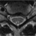
87 Carpal Tunnel Disease and Wrist Fractures The small structures involved in wrist MRI necessitate maximization of spatial resolution and SNR with dedicated surface coils and imaging at 3 T. The effect of such measures is illustrated by the ability to detect injuries to small structures like the median nerve, which is affected in carpal tunnel syndrome, illustrated in Fig. 87.1. Here (A) axial T1WI and (B) FS T2WI demonstrate enlargement of the nerve as it courses between flexor tendons deep to the flexor retinaculum at the level of the hamate. Calculating ratios of ipsilateral nerve diameters at the pisiform or hamate versus at the distal radius may prove more useful in documenting enlargement than contralateral size comparisons, as carpal tunnel syndrome is bilateral in more than half of cases. Figure 87.1B
![]()
Stay updated, free articles. Join our Telegram channel

Full access? Get Clinical Tree


