46 Cerebral Arterial Circle (Circle of Willis)
F. Goetz, A. Giesemann
There are many anomalies of the arteries at the base of the skull.1–7 Some studies deal with anomalies of the internal carotid artery8–13 and others with the circle of Willis14–26 or its branches as the middle cerebral artery27–30 or cerebellar arteries.31,32 For didactic reasons, the anomalies of the anterior area,19,33–36 posterior area,19,37–39 and the basilar artery40 are depicted separately. These anomalies can be combined in different individuals and result in many different types. The frequencies given here are a compromise between radiological and anatomical studies. Vessels were termed “small” or “absent” (e.g., Fig. 46.13) when only negligible collateral circulation took place via such a route.
The recognition of cerebral variants is important as some of these variants might turn pathological in a surgical setting and can influence pathological changes such as brain infarcts and aneurysms.7
46.1 Anterior Part of the Cerebral Circle
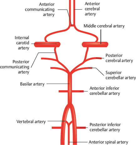
Fig. 46.1 Circle of Willis. Schematic with indication of arteries.

Fig. 46.2 “Normal” situation (60%). Schematic.

Fig. 46.3 Two anterior communicating arteries (10%).

Fig. 46.4 The median artery of the corpus callosum originates from the anterior communicating artery (10%). Schematic.

Fig. 46.5 Instead of the anterior communicating artery, a lateral anastomosis between the two anterior cerebral arteries is present (3%). Schematic (a) and X-ray angiography, anterior view of the right and left internal carotid artery (b). 1 Anterior communicating artery; 2 anterior cerebral artery; 3 middle cerebral artery; 4 internal carotid artery.

Fig. 46.6 Both anterior cerebral arteries combine partly in a common trunk (5%). Schematic.

Fig. 46.7 Both anterior cerebral arteries originate from one internal carotid artery (10%). Schematic.

Fig. 46.8 Absence of the anterior communicating artery (1%). Schematic.

Fig. 46.9 Accessory middle cerebral artery (1%). Schematic.

Fig. 46.10 Absence of one internal carotid artery; the middle cerebral artery originates from the basilar artery (0.1%). Schematic.

Fig. 46.11 Absence of one internal carotid artery; the middle cerebral artery originates from the other internal carotid artery (0.1%). Schematic.
46.2 Posterior Part of the Cerebral Circle

Fig. 46.12 “Normal situation (55%). Schematic.

Fig. 46.13 The posterior communicating artery is absent or very small on one side (10%). Schematic.

Fig. 46.14 The posterior communicating artery is absent or very small on both sides (<1%). Schematic.

Fig. 46.15 On one side the posterior cerebral artery originates from the internal carotid artery (<1%). Schematic (a) and X-ray angiography, right internal carotid artery injection, lateral view with a dominant posterior communicating artery and anterior view of the left vertebral artery; not hypoplasia of the so-called P1-segment of the right posterior cerebral artery (b). 1 Posterior cerebral artery (P2 segment); 2 vertebral artery; 3 basilar artery; 4 right internal carotid artery; 5 posterior communicating artery.
Related posts:


Stay updated, free articles. Join our Telegram channel

Full access? Get Clinical Tree


