• Asymmetric hyperextension pattern of injury to posterior column
• Below C2; most common at C6 and C7
• Fracture of articular facet
Associated with radiculopathy
• Fracture of articular pillar
• Simultaneous fractures of pedicle and ipsilateral lamina = traumatic isolation of cervical articular pillar
• May see degree of rotational deformity at level of injury
• Best imaging modality
Thin slice (≤ 1 mm) helical CT with sagittal and coronal reformations
MR (especially STIR) to evaluate ligaments, spinal cord
• Radiographs should include oblique views to evaluate facet alignment, articular pillars
Top Differential Diagnoses
• Hyperflexion injury, cervical
• Hyperflexion-rotation injury, cervical
• Lateral flexion injury, cervical
Pathology
• Rotational instability
• Radiculopathy
Clinical Issues
• Surgical fusion to achieve rotational stability
(Left) Axial NECT shows unilateral fractures of the right C6 articular pillar extending into the lamina .
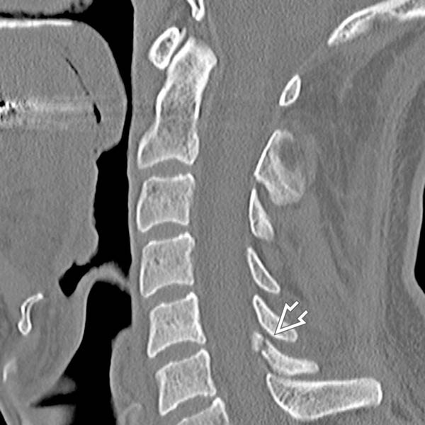
(Right) Sagittal reformatted CT in the same patient shows extension of the articular pillar fracture into the ipsilateral lamina .
(Left) Coronal reconstructed CT of the hyperextension-rotation injury shows a unilateral comminuted, mildly displaced fracture involving the right C7 articular pillar .
(Right) Axial NECT shows fractures of the right C6 articular pillar and lamina , resulting in traumatic isolation of the articular pillar.
TERMINOLOGY
Synonyms
• Pedicolaminar fracture-separation
Definitions
• Hyperextension injury to cervical spine with off-center force vector causing rotation and asymmetric injury
IMAGING
General Features
• Best diagnostic clue
Asymmetric hyperextension pattern of injury to posterior column
• Location
Below C2; most common at C6 and C7
• Fracture of articular facet
Simple fracture or impacted/comminuted
Fracture of superior articular facet more commonly associated with radiculopathy
Only gold members can continue reading. Log In or Register to continue


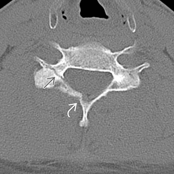
 extending into the lamina
  .
.
 .
.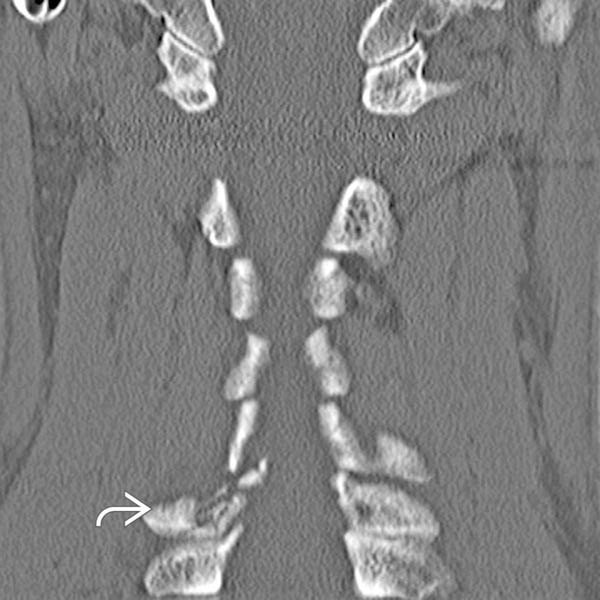
 .
.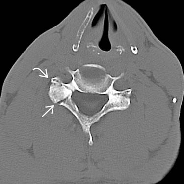
 and lamina
  , resulting in traumatic isolation of the articular pillar.


