Chapter 30 Cervical Spinal Cord Stimulation
Note: Please see page ii for a list of anatomical terms/abbreviations used in this book.
Access to the cervical interlaminar space may be obtained from between C7-T1 through T2-T3.
 Trajectory View (Figure 30–1)
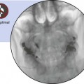
 Confirm the appropriate interlaminar space with the anteroposterior view. (We demonstrate T1-2 here.)


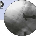
 Remove the 18-gauge needle, and use the introducer needle to obtain the trajectory needle view.
 Because this is the trajectory view, the needle entry position should be parallel to the C-arm beam.

Related posts:
 Atlantoaxial Joint Intraarticular Injection
 Cervical Zygapophysial Joint Nerve (Medial Branch) Radiofrequency Neurotomy and Nerve Injection, Posterior Approach
Stay updated, free articles. Join our Telegram channel

Full access? Get Clinical Tree


