•Pneumothorax
°Tension pneumothorax (idiopathic, traumatic pneumothorax with valve mechanism, etc.)
°Pneumothorax on mechanical ventilation
°Persistent or recurrent iatrogenic pneumothorax after needle aspiration
°Symptomatic pneumothorax
°Large pneumothorax
•Malignant pleural effusion
•Empyema (stage-dependent) and complicated parapneumonic effusions
•Traumatic hemothorax secondary to chest trauma
•Post-operative care (e.g. after thoracotomy, video-assisted thoracoscopy, coronary bypass)
Physiology in the Pleural Space
The pleural space is defined by the space between the visceral and parietal pleura, which contains a small amount of fluid generating a mechanical connection of the lung with the chest wall, due to their passive and elastic structure.
The intrapleural pressure alternates during a breathing cycle in between −8 cm H20 (inspiration) and −2 cm H20 (expiration). Forced inspiration and expiration lead to a pressure difference of −54 cm H20 to + 70 cm H20. However, the negative intrapleural pressure cannot be sustained when fluid or gas enters the pleural space. That, in the absence of pleural adhesions, results in a collapse of the lung with hypoxemia and alveolar hypoventilation. If the pressure in the pleural space increases to a tension pneumothorax, shifting of the mediastinum to the contralateral lung and decrease venous flow of blood back to the heart can result in severe hypoxemia and hemodynamic collapse. By insertion of a chest tube, draining of the pleural space and restoration of the physiologic pressure conditions can be obtained.
Indications for Chest Tubes
The initial therapy for every symptomatic pneumothorax is the immediate placement of a tube. Diagnostic workup has to be postponed when clinical symptoms and signs of pneumothorax start to be life threatening. One exclusion to this rule is selected cases with primary (idiopathic) spontaneous pneumothorax (PSP) or asymptomatic partial PSP (pneumothorax, with a pleural separation < 1 cm). These patients can be observed under close surveillance. In an emergency with a symptomatic tension pneumothorax, when a regular chest tube placement cannot be achieved in time, intercostal puncture with a wide lumen indwelling catheter can result in a temporary release of the life-threatening situation. In patients under mechanical ventilation, developing pneumothoraces make chest tube placement almost always mandatory. These patients can quickly establish a tension pneumothorax.
In iatrogenic pneumothorax, after transbronchial biopsy, transthoracic needle aspiration or paravertebral nerve blocks (pain therapy) or after puncture of the V. subclavia (central venous catheter), a lung parenchyma injury with pneumothorax may develop. After every procedure, a chest X-ray should be performed. After minor interventions, asymptomatic pneumothoraces with small apical separations can be frequently observed. In these patients, close observation is recommended as these pneumothoraces can progress.
In patients with hemothorax, it is essential to evaluate the degree of bleeding. Inserting a chest tube can facilitate re-expansion of the lung and may avoid trapping of the lung as well as late empyema development.
The situation of parapneumonic fluid collection is controversial. Does it need a tube placement? Commonly accepted is the tube placement in stage II empyema (ATS-Classification 1962). Tube placement in a stage I empyema with residual fluid or not fully expanded lung can help drain the pleural cavity and assist full lung expansion.
In symptomatic or recurrent malignant pleural effusion, an insertion of a chest tube is indicated because of diagnostic and palliative reasons, as well as in cases in which a pleurodesis is considered.
Contraindications for Chest Tube Insertion
Absolute contraindications do not exist. Relative contraindications are found in patients with bleeding disorders or anticoagulation therapy. Special conditions include pleural adhesions, loculated pleural effusions or empyema, pulmonary giant bullae – misinterpreted as pneumothorax – and, in trauma patients, rupture of the diaphragm with thoracic displacement of intra-abdominal organs. Under these circumstances, computed tomography or ultrasound should be applied, and the operating room should be in standby.
Patient Consent
The patient has to be informed about indication, technique and impact on their health condition. The use of local anaesthesia as well as the possibility for additional sedation should be mentioned. In addition to the general information sheet, every possible complication should be explained and described, and especially the following ones should be mentioned:
Improper placement
Tube dislodgement
Organ penetration with bleeding, bronchopleural fistula
Empyema – chest tube placement could introduce bacteria into the pleural space
Re-expansion (oedema) of the lung with coughing, shoulder and thoracic pain as well as vagal reactions
Injury to the intercostal blood vessels/nerves and the periosteum of the rib with bleeding, pain and intercostal neuralgia
Injury of intraperitoneal or intrathoracic organs
Emergency thoracotomy
Need for additional procedures
Sizing of Chest Tubes on the Basis of Indication
Appropriate are silicone tubes in a size between 6 and 32 French with length marking, contrast line and multiple side perforations. Straight and right-angled tubes are available. The size of the tube that is needed depends on the indication for the chest tube insertion (recommended sizes for pneumothorax are 20 Fr, 24–28 Fr for effusion), as well as considerations for gender and size of the patient.
Preoperative Diagnostic Workup
The insertion of the chest tube has to be done after accurate clinical examination and after review of the X-ray, chest CT or ultrasound. The only exclusion is the urgent, clinical suspicion of a tension pneumothorax with loss of blood pressure, hypoxemia, tachypnoea, superior vena cava syndrome and high ventilation pressures.
Pleural Chest Tube Insertion
Position of the Patient, Anatomy, Anaesthesia and Technique
The insertion of the chest tube has to be done under sterile conditions with special instruments (Fig. 57.1). In the situation of non-loculated processes, the insertion should be done in supine position or at an angle of 45°; the arm on the affected side should be abducted and externally rotated.
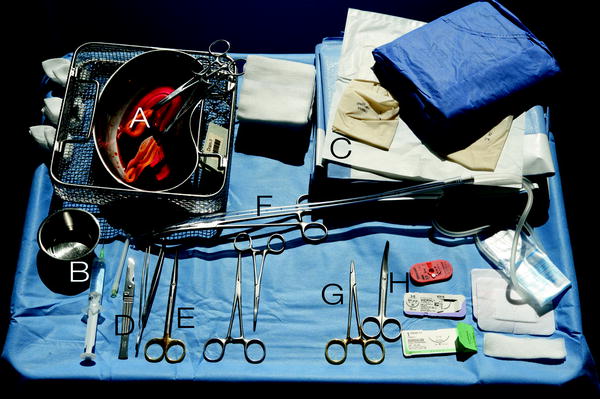

Fig. 57.1
Instruments: (a) Disinfectant, (b) Local anaesthesia, (c) Sterile drapes, (d) Scalpel and forceps, (e) Scissors, (f) Pleural drainage and Kelly clamp, (g) Needle clamp, (h) Scissors and suture
Stay updated, free articles. Join our Telegram channel

Full access? Get Clinical Tree


