Table 5.98 Child abuse: conditions that masquerade as child abuse and simulate the classic metaphyseal lesion (see Table 5.44)
Diagnosis
Comments
Birth trauma
After low segment cesarean sections.
Iatrogenic
After orthopedic manipulations.
Osteomyelitis
Osteomyelitis has a predilection for the metaphysis in infants. Although systemic signs and symptoms of infection may be lacking in infants and multiple sites of osteomyelitis and different stages of healing may simulate child abuse, the metaphyseal erosions in osteomyelitis tend to be less well-defined than classic metaphyseal lesions (CMLs; see Table 5.43).
Controversy exists as to whether or not osseous fragments resembling CMLs are always accompanied by more obvious changes of rickets such as metaphyseal irregularity and physeal widening.70,71 Laboratory findings may also help support a diagnosis of rickets.
Bone dysplasias
Skeletal survey will reveal other abnormalities associated with the given skeletal dysplasia. DD: osteogenesis imperfecta, metaphyseal chondrodysplasia (Schmid type), and spondylometaphyseal dysplasia (corner fracture type).
Leukemia
Bone demineralization, osteolytic lesions, and subperiosteal new bone formation. Radiolucent bands at the metaphyses. Imaging findings may be due to a combination of nutritional disturbance and leukemic invasion (see Table 5.43).
Scurvy
Subperiosteal hemorrhage and metaphyseal and epiphyseal changes. Characteristic findings of scurvy may help to distinguish from child abuse (see Table 5.43).
Vitamin A intoxication
Subperiosteal new bone formation predominantly in the tubular bones.
Lower extremity paralysis
Routine handling of infants with paralysis may cause fractures.
Infantile cortical hyperostosis (Caffey disease)
Subperiosteal new bone formation and cortical thickening. Preference for the mandible, clavicle, and ulna.
Menkes syndrome (kinky hair disease)
Rare genetic disorder resulting in defective gastrointestinal absorption of copper. Metaphysis of the long bones have spurs ± fractures. Subperiosteal new bone formation and bone demineralization. Tortuous and irregular cerebral and abdominal arteries.
Congenital indifference/insensitivity to pain
Lack of sensation of pain may lead to a delay in diagnosis of both minor and major trauma. Imaging may show metaphyseal injuries, subperiosteal hemorrhage, fractures, and epiphyseal separations in various stages of healing during infancy. Charcot-type joints manifest later in childhood.
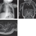
Fig. 5.180 Rickets and metabolic bone disease. Metaphyseal irregularity simulating a metaphyseal corner fracture (arrow) in a 3-year-old girl with resorptive metabolic bone disease of unclear etiology.
Table 5.99 Child abuse: specificity of fracture locations
Table 5.99 reproduced with permission from Kleinman PK. Diagnostic imaging of child abuse. 2nd ed. St. Louis: Mosby, 1989.
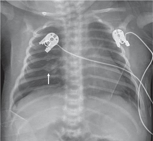
Fig. 5.181 Healing posterior rib fractures (arrow) in child abuse.
References
1. Restrepo CS, Martinez S, Lemos DF, et al. Imaging appearances of the sternum and sternoclavicular joints. Radiographics 2009;29:839–8592. Yekeler E, Tunaci M, Tunaci A, Dursun M, Acunas G. Frequency of sternal variations and anomalies evaluated by MDCT. AJR Am J Roentgenol 2006;186:956–9603. Jeung MY, Gangi A, Gasser B, et al. Imaging of chest wall disorders. Radiographics 1999;19:617–6374. Edwards DK 3rd, Berry CC, Hilton SW. Trisomy 21 in newborn infants: chest radiographic diagnosis. Radiology 1988;167: 317–3185. Levine MS, Borden S 4th, Gill FM. Sternal cupping: a new finding in childhood sickle cell anemia. Radiology 1982;142:367–3706. Mehta AV, Chidambaram B, Suchedina AA, Garrett AR. Radiologic abnormalities of the sternum in Turner’s syndrome. Chest 1993;104:1795–17997. Khanna G, Sato TS, Ferguson P. Imaging of chronic recurrent multifocal osteomyelitis. Radiographics 2009;29:1159–11778. Kimonis VE, Mehta SG, Digiovanna JJ, Bale SJ, Pastakia B. Radiological features in 82 patients with nevoid basal cell carcinoma (NBCC or Gorlin) syndrome. Genet Med 2004;6:495–5029. Glass RB, Norton KI, Mitre SA, Kang E. Pediatric ribs: a spectrum of abnormalities. Radiographics 2002;22:87–10410. Kumar R, Madewell JE, Swischuk LE, Lindell MM, David R. The clavicle: normal and abnormal. Radiographics 1989;9:677–70611. Oestreich AE. The lateral clavicle hook-an acquired as well as a congenital anomaly. Pediatr Radiol 1981;11:147–15012. Lachman R. Taybi and Lachman’s radiology of syndromes, metabolic disorders and skeletal dysplasias. 5th ed. St. Louis: Mosby, 200613. OMIM: Online Mendelian Inheritance in Man Web site. Johns Hopkins University. http://www-ncbi-nlm-nih-gov.easyaccess1.lib.cuhk.edu.hk/omim. Accessed July 200914. Waters PM, Smith GR, Jaramillo D. Glenohumeral deformity secondary to brachial plexus birth palsy. J Bone Joint Surg Am 1998;80:668–67715. Wetherell RG, Amis AA, Heatley FW. Measurement of acetabular erosion. The effect of pelvic rotation on common landmarks. J Bone Joint Surg Br 1989;71:447–45116. Paajanen H, Hermunen H, Karonen J. Pubic magnetic resonance imaging findings in surgically and conservatively treated athletes with osteitis pubis compared to asymptomatic athletes during heavy training. Am J Sports Med 2008;36: 117–12117. Muecke EC, Currarino G, Currarino G. Congenital widening of the pubic symphysis: associated clinical disorders and roentgen anatomy of affected bony pelves. Am J Roentgenol Radium Ther Nucl Med 1968;103:179–18518. Zook PD, Winter TC 3rd, Nyberg DA. Iliac angle as a marker for Down syndrome in second-trimester fetuses: CT measurement. Radiology 1999;211:447–45119. Klein DM, Barbera C, Gray ST, Spero CR, Perrier G, Teicher JL. Sensitivity of objective parameters in the diagnosis of pediatric septic hips. Clin Orthop Relat Res 1997:153–15920. Simmons BP, Southmayd WW, Riseborough EJ. Congenital radioulnar synostosis. J Hand Surg Am 1983;8:829–83821. Steinberg ME, Steinberg DR. Classification systems for osteonecrosis: an overview. Orthop Clin North Am 2004;35:273–283, vii–viii22. Laor T, Jaramillo D. MR imaging insights into skeletal maturation: what is normal? Radiology 2009;250:28–3823. Gardner DJ, Azouz EM. Solitary lucent epiphyseal lesions in children. Skeletal Radiol 1988;17:497–50424. Varich LJ, Laor T, Jaramillo D. Normal maturation of the distal femoral epiphyseal cartilage: age-related changes at MR imaging. Radiology 2000;214:705–70925. Jaramillo D, Shapiro F, Hoffer FA, et al. Posttraumatic growth-plate abnormalities: MR imaging of bony-bridge formation in rabbits. Radiology 1990;175:767–77326. Levesque M, Legmann P, Le Cloirec A, Deybach JC, Nordmann Y. Radiological features in congenital erythropoietic porphyria (Gunther’s disease). Review of 3 cases. Pediatr Radiol 1988;18:62–6627. Kleinman PK. Problems in the diagnosis of metaphyseal fractures. Pediatr Radiol 2008;38(Suppl 3):S388–S39428. Kleinman PK. Diagnostic imaging of child abuse. 2nd ed. St. Louis: Mosby, 198929. Grayev AM, Boal DK, Wallach DM, Segal LS. Metaphyseal fractures mimicking abuse during treatment for clubfoot. Pediatr Radiol 2001;31:559–56330. Kozlowski K, Sutcliffe J, Barylak A, et al. Hypophosphatasia. Review of 24 cases. Pediatr Radiol 1976;5:103–11731. Goldfarb CA, Manske PR, Busa R, Mills J, Carter P, Ezaki M. Upper-extremity phocomelia reexamined: a longitudinal dysplasia. J Bone Joint Surg Am 2005;87:2639–264832. Le Goff C, Cormier-Daire V. Genetic and molecular aspects of acromelic dysplasia. Pediatr Endocrinol Rev 2009;6:418–42333. Diren HB, Kutluk MT, Karabent A, Gocmen A, Adalioglu G, Kenanoglu A. Primary hypertrophic osteoarthropathy. Pediatr Radiol 1986;16:231–23434. Cheema JI, Grissom LE, Harcke HT. Radiographic characteristics of lower-extremity bowing in children. Radiographics 2003;23:871–88035. Nguyen ML, Jones NF. Undergrowth: brachydactyly. Hand Clin 2009;25:247–25536. Temtamy SA, Aglan MS. Brachydactyly. Orphanet J Rare Dis 2008;3:1537. Hussmann J, Russell RC, Kucan JO, Khardori R, Steinau HU. Soft-tissue calcifications: differential diagnosis and therapeutic approaches. Ann Plast Surg 1995;34:138–14738. Wong KS, Chiu CH, Huang YC, Lin TY. Childhood and adolescent tuberculosis in northern Taiwan: an institutional experience during 1994–1999. Acta Paediatr 2001;90:943–94739. Houser JR, Kan JH. Langerhans cell histiocytosis of the epiphysis. Pediatr Radiol 2008;38:8551840. Brien EW, Mirra JM, Ippolito V, Vaughan L. Clear-cell chondrosarcoma with elevated alkaline phosphatase, mistaken for osteosarcoma on biopsy. Skeletal Radiol 1996;25:770–77441. Resnick D, Greenway G. Distal femoral cortical defects, irregularities, and excavations. Radiology 1982;143:345–35442. Mar WA, Taljanovic MS, Bagatell R, et al. Update on imaging and treatment of Ewing sarcoma family tumors: what the radiologist needs to know. J Comput Assist Tomogr 2008;32:108–11843. Gladish GW, Sabloff BM, Munden RF, Truong MT, Erasmus JJ, Chasen MH. Primary thoracic sarcomas. Radiographics 2002;22:621–63744. Ablin DS, Azouz EM, Jain KA. Large intrathoracic tumors in children: imaging findings. AJR Am J Roentgenol 1995;165:925–93445. Bonetumor.org Web site. bonetumor.org. Accessed July 30, 200946. Greenspan A, Jundt G, Remagen W. Differential diagnosis in orthopaedic oncology. 2nd ed. Philadelphia: Lippincott Williams & Wilkins, 200747. Moppett J, Oakhill A, Duncan AW. Second malignancies in children: the usual suspects? Eur J Radiol 2001;38:235–24848. Gartner L, Pearce CJ, Saifuddin A. The role of the plain radiograph in the characterisation of soft tissue tumours. Skeletal Radiol 2009;38:549–55849. Van Rijn RR, Wilde JC, Bras J, Oldenburger F, McHugh KM, Merks JH. Imaging findings in noncraniofacial childhood rhabdomyosarcoma. Pediatr Radiol 2008;38:617–63450. Beall DP, Ly J, Bell JP, Parker EE, et al. Pediatric extraskeletal osteosarcoma. Pediatr Radiol 2008;38:579–58251. McCarville MB, Kaste SC, Pappo AS. Soft-tissue malignancies in infancy. AJR Am J Roentgenol 1999;173:973–97752. Garces-Inigo EF, Leung R, Sebire NJ, McHugh K. Extrarenal rhabdoid tumours outside the central nervous system in infancy. Pediatr Radiol 2009;39:817–82253. Subbarao K. Periosteal reactions in pediatrics. Indian J Pediatr 1987;54:45–5254. Makley JT, Dunn MJ. Prostaglandin synthesis by osteoid osteoma. Lancet 1982;2:4255. Poznanski AK, Fernbach SK, Berry TE. Bone changes from prostaglandin therapy. Skeletal Radiol 1985;14:20–2556. Shabshin N, Schweitzer ME, Morrison WB, Carrino JA, Keller MS, Grissom LE. High-signal T2 changes of the bone marrow of the foot and ankle in children: red marrow or traumatic changes? Pediatr Radiol 2006;36:670–67657. Zubler V, Mengiardi B, Pfirrmann CW, et al. Bone marrow changes on STIR MR images of asymptomatic feet and ankles. Eur Radiol 2007;17:3066–307258. McQueen FM. Magnetic resonance imaging in early inflammatory arthritis: what is its role? Rheumatology (Oxford) 2000;39:700–70659. Mora S, Gilsanz V. Establishment of peak bone mass. Endocrinol Metab Clin North Am 2003;32:39–6360. Gilsanz V, Perez FJ, Campbell PP, Dorey FJ, Lee DC, Wren TA. Quantitative CT reference values for vertebral trabecular bone density in children and young adults. Radiology 2009;250:222–22761. Cohen MM Jr. A comprehensive and critical assessment of over-growth and overgrowth syndromes. Adv Hum Genet 1989;18: 181–303, 373–37662. Green DM, Breslow NE, Breslow NE, Beckwith JB, Norkool P. Screening of children with hemihypertrophy, aniridia, and Beck-with-Wiedemann. Med Pediatr Oncol 1993;21:188–19263. Goldman AB, Kaye JJ. Macrodystrophia lipomatosa: radiographic diagnosis. AJR Am J Roentgenol 1977;128:101–10564. Spranger J. Radiologic nosology of bone dysplasias. Am J Med Genet 1989;34:96–10465. Patel MD, Filly RA. Homozygous achondroplasia: US distinction between homozygous, heterozygous, and unaffected fetuses in the second trimester. Radiology 1995;196:541–54566. Prosser I, Maguire S, Harrison SK, Mann M, Sibert JR, Kemp AM. How old is this fracture? Radiologic dating of fractures in children: a systematic review. AJR Am J Roentgenol 2005;184:1282–128667. Offiah A, van Rijn RR, Perez-Rossello JM, Kleinman PK. Skeletal imaging of child abuse (non-accidental injury). Pediatr Radiol 2009;39:461–47068. O’Connor JF, Cohen J. Dating fractures. In: Kleinman PK, ed. Diagnostic imaging of child abuse. 2nd ed. St. Louis: Mosby, 1998:168–17769. Brill PW, Winchester P, Kleinman PK. Differential diagnosis I: diseases simulating abuse. In: Kleinman PK, ed. Diagnostic imaging of child abuse. 2nd ed. St. Louis: Mosby, 1998:178–19670. Keller KA, Barnes PD. Rickets vs. abuse: a national and international epidemic. Pediatr Radiol 2008;38:1210–121671. Slovis TL, Chapman S. Evaluating the data concerning vitamin D insufficiency/deficiency and child abuse. Pediatr Radiol 2008;38:1221–122472. Kleinman PK. Skeletal trauma: general considerations. In: Klein-man PK, ed. Diagnostic imaging of child abuse. 2nd ed. St. Louis: Mosby, 1998:168–177
Only gold members can continue reading. Log In or Register to continue