CHAPTER 8 Cholesteatoma of the External Auditory Canal
Epidemiology
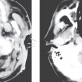
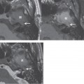
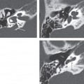
External auditory canal cholesteatomas (EACC) are rare, with an incidence of 1 in 1000 new otologic patients. Their exact etiology is unknown, and many may occur spontaneously, although prior inflammation, trauma, or surgery have all been associated with EACC. EACC may be caused by loss of normal lateral epithelial migration from the tympanic membrane and external auditory canal (EAC); or increased cellular proliferation induced by accumulation of keratin debris may play a role in the development of these lesions. EAC stenosis or obstruction may also result in EACC. Although EACC can develop in any portion of the canal, the posterior-inferior wall is most common.
Clinical Features
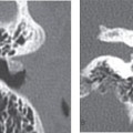
Most patients are adults presenting between 40 and 75 years of age with otorrhea and dull otalgia. Rarely, hearing loss may occur secondary to obstruction of the canal by the EACC. Facial nerve paralysis is even less common. Examination reveals squamous debris protruding from the involved canal wall, which unlike keratosis obturans, does not form a plug. Purulent drainage may be present deeper in the canal due to associated infection. Localized bony erosion is usually present and visible after removal of Chol the debris. Clinically it may appear similar to keratosis obturans, postinflammatory medial canal fibrosis, malignant external otitis, and squamous cell carcinoma.
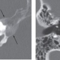
Pathology
Related posts:

Stay updated, free articles. Join our Telegram channel

Full access? Get Clinical Tree


