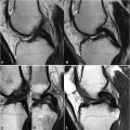
80 Collateral Ligaments, Knee Like other ligaments, type 1 collagen lends the collateral knee ligaments of the knee a hypointense appearance on common pulse sequences. The superficial portion of the MCL arises from the medial femoral condyle and inserts below the joint line, merging with low SI cortical bone posterior to the pes anserinus muscles. The course of this ligament is illustrated on the coronal FS and non-FS PDWI of Figs. 80.1A,B, respectively. The superficial MCL fibers are separated from the deep fibers by the Voshell bursa. The pes anserinus bursa lies distal to this, anterior to the tendons of the sartorius, semitendinosus, and gracilis. The deep MCL is best seen in the PDWI of Fig. 80.1A, merging with the joint capsule and medial meniscus medial to the superficial portion. Grade 1 lesions (i.e., sprain) demonstrate edema and/or hemorrhagic SI in the tissues superficial to the MCL with the ligament itself remaining normal in SI and thickness. Grade 2 lesions are partial tears marked by displacement from adjacent bone with intraligamentous hyperintensity on STIR and FS PDWI often present. A grade 3 lesion is a complete tear and is illustrated on the coronal FS T2 and PDWI in Figs. 80.1C,D. Here, particularly on the (C) FS T2WI, thickening and high SI edema within the MCL with full-thickness disruption (black arrow) of the proximal fibers of the superficial and deep ligamentous components are visualized. These features are not as clearly demonstrated on the (D) PDWI without fat saturation, although compared with a normal MCL (Fig. 80.1B
![]()
Stay updated, free articles. Join our Telegram channel

Full access? Get Clinical Tree


