A. No intervention needed, the study is normal
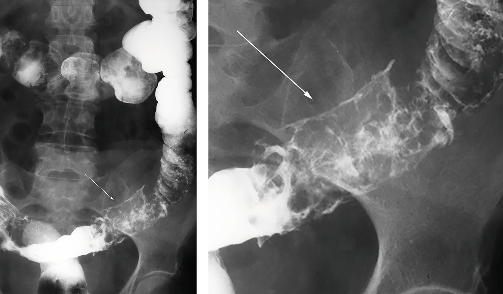
B. Diet modification with course of oral broad-spectrum antibiotics
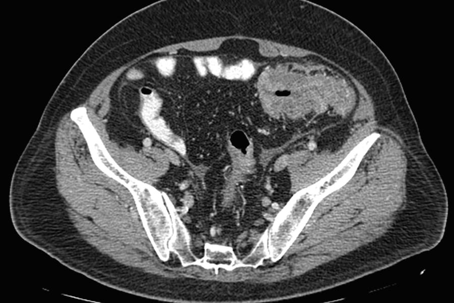
C. Colonoscopy to evaluate for suspected malignancy
D. Urgent surgical consultation
E. Single-contrast enema to assess for degree of obstruction
A. Acute diverticulitis is the most common cause of large bowel obstruction.
B. The diverticula are traction-type pseudodiverticula caused by adjacent fibrosis.
C. Diverticula are most common in the rectum and sigmoid colon.
D. Diverticula form at the site of the vasa recta penetration of the bowel wall.
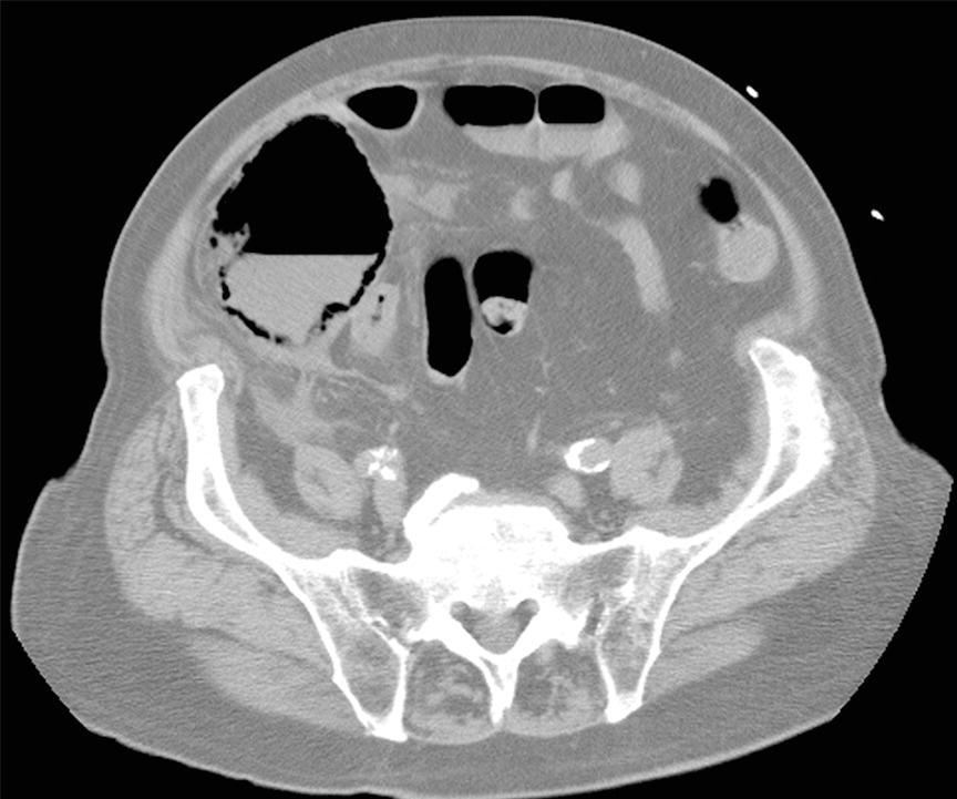
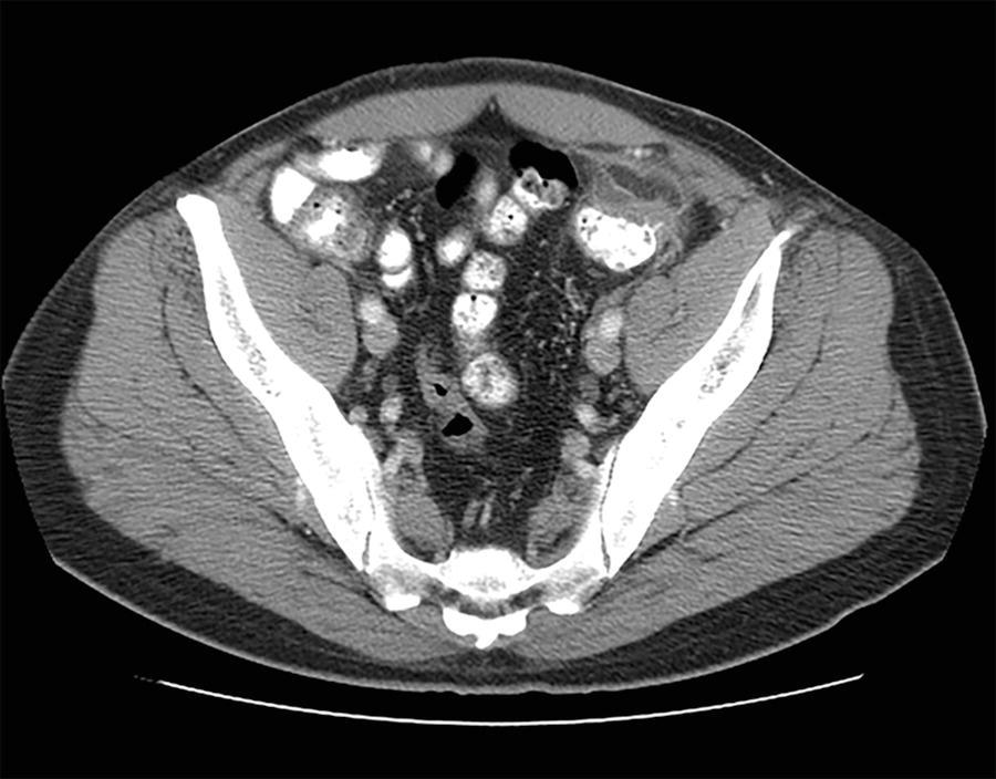
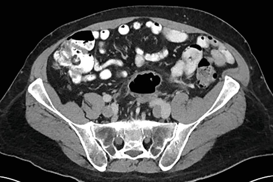
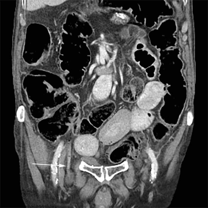
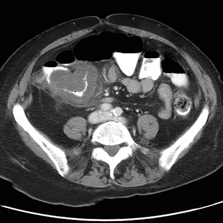
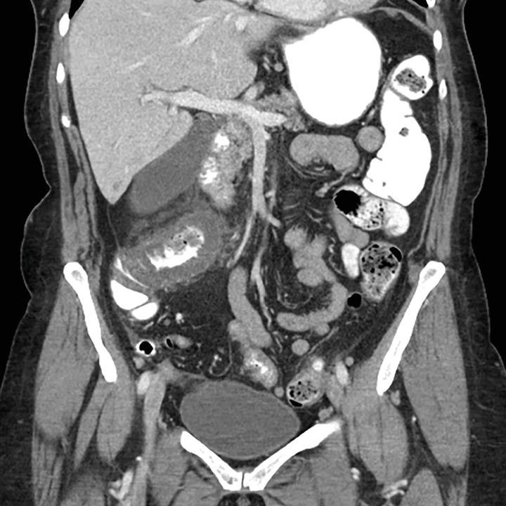
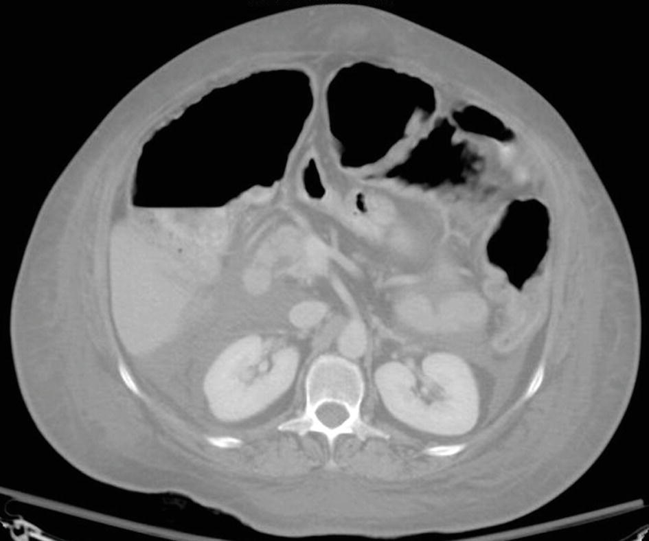
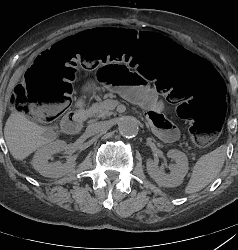
2 A 34-year-old female presents with 2 days onset of right lower quadrant pain. On examination, the patient has a mild leukocytosis and low-grade fever. A CT performed with intravenous contrast was obtained. What is the most appropriate next step?
A. Pelvic ultrasound to assess for tuboovarian abscess
B. Repeating the CT study using enteric contrast
C. Surgical consultation
D. No treatment needed for a self-limited condition
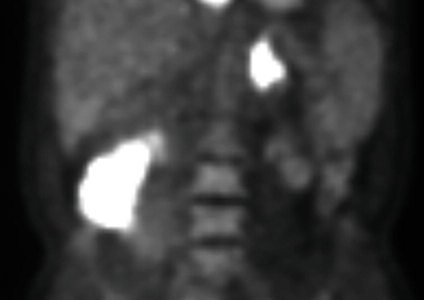
3a A 70-year-old female with a history of coronary artery disease and prior myocardial infarcts presents with rectal bleeding and severe abdominal pain. A contrast-enhanced CT scan and follow-up barium enema demonstrates a finding in the distal transverse colon. What is the most likely etiology?

A. Crohn disease
B. Radiation colitis
C. Ischemic colitis
D. Chronic diverticulitis
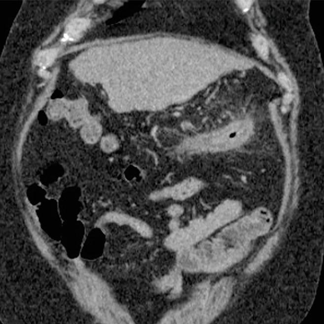
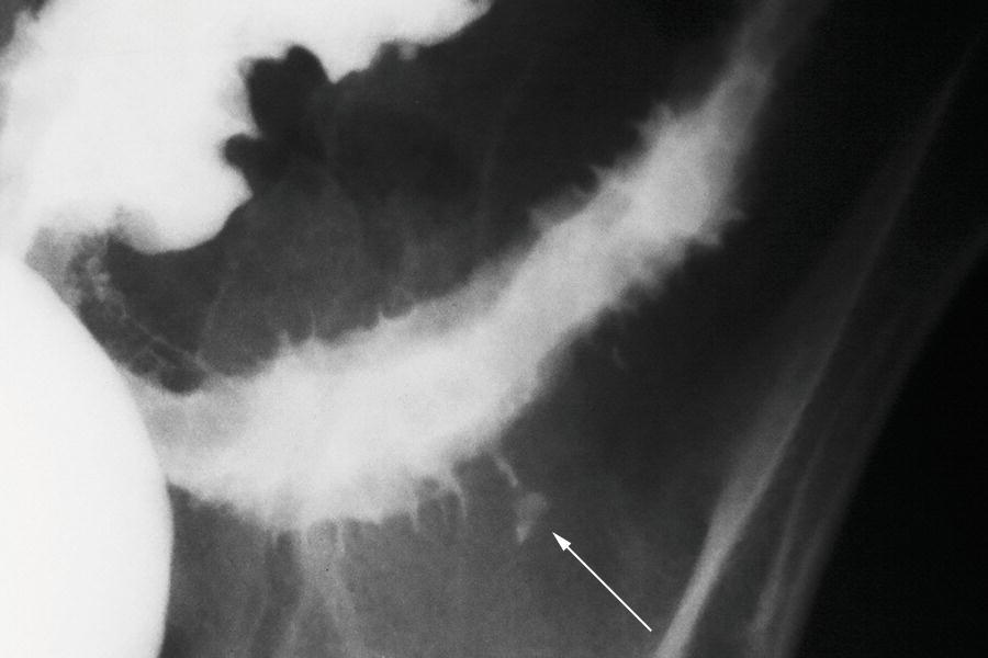
3b What is the name of the watershed area in the colon between the superior and inferior mesenteric arterial territories?
A. Meyers point
B. Drummond point
C. Sudeck point
D. Griffiths point
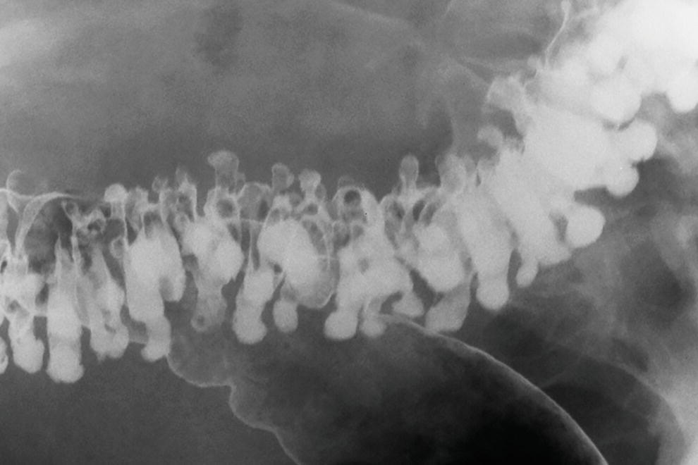
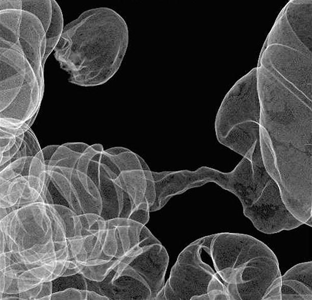
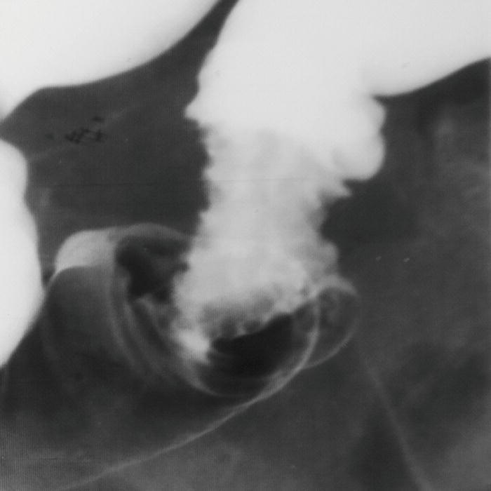
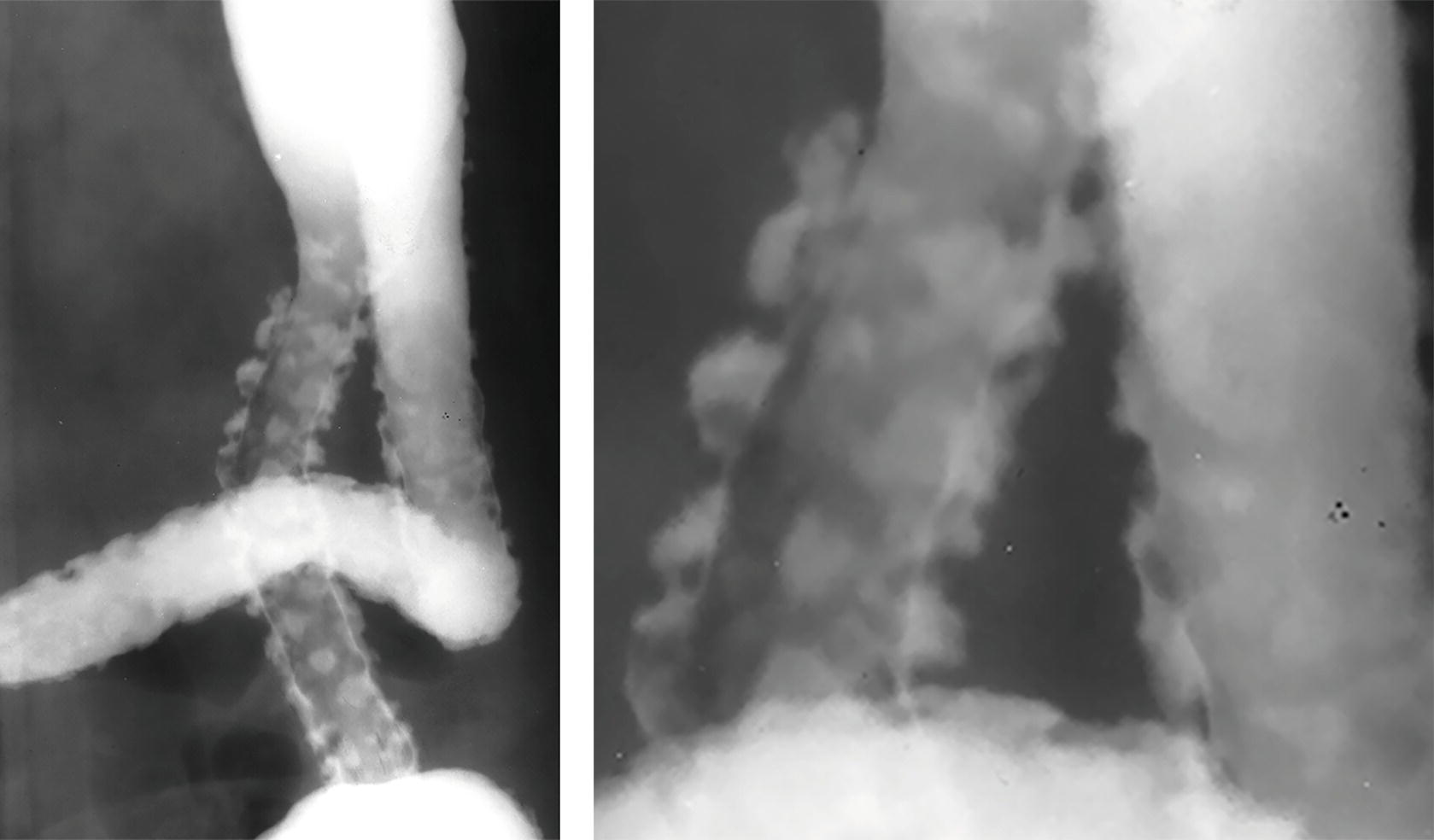
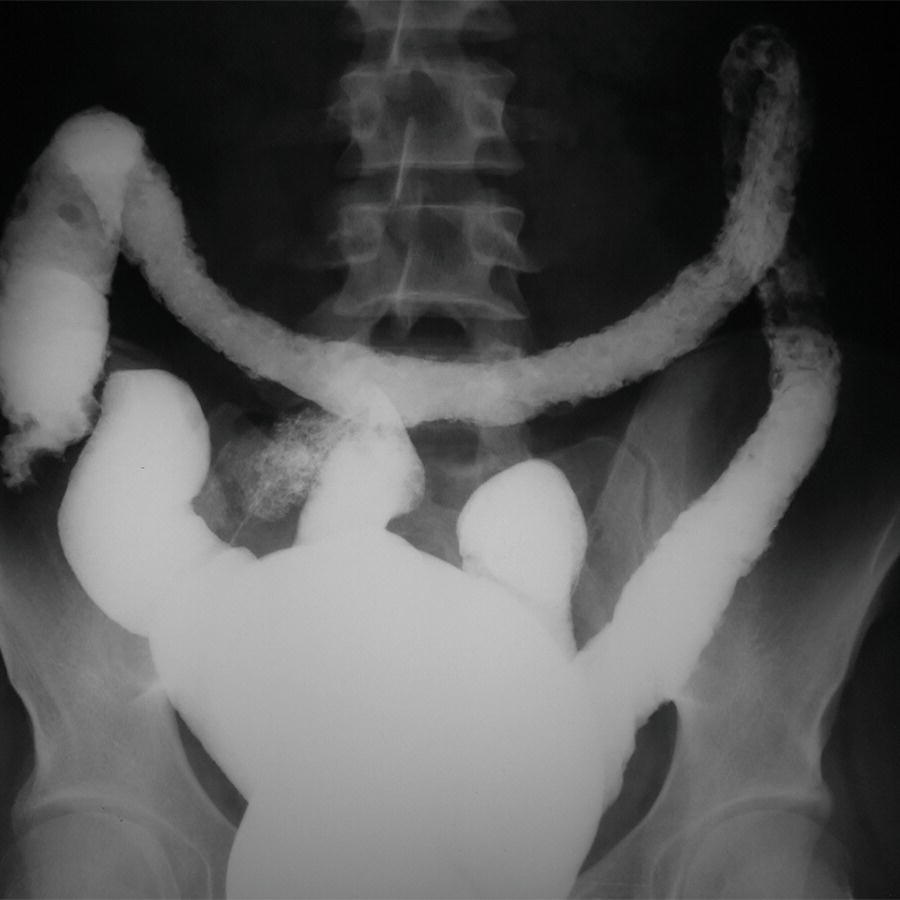
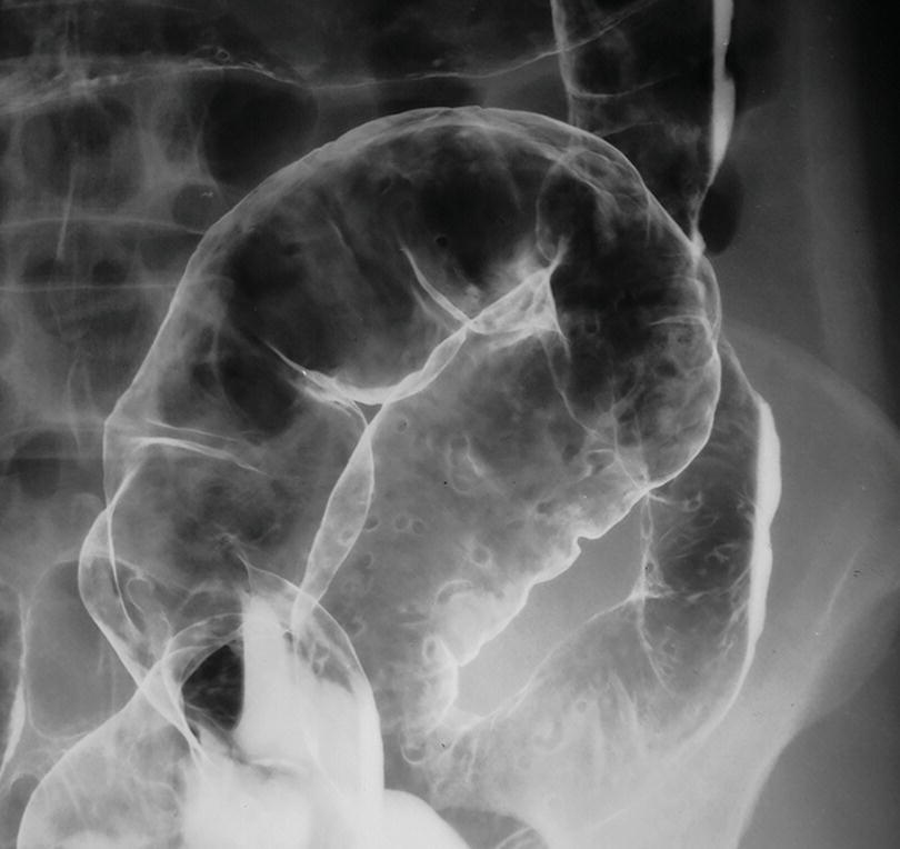
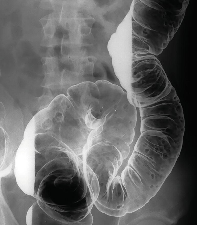
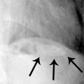
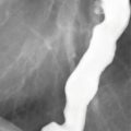
4 A single-contrast barium enema is performed on a 50-year-old male. A spot film of the sigmoid colon is shown below. What is the appropriate next step?
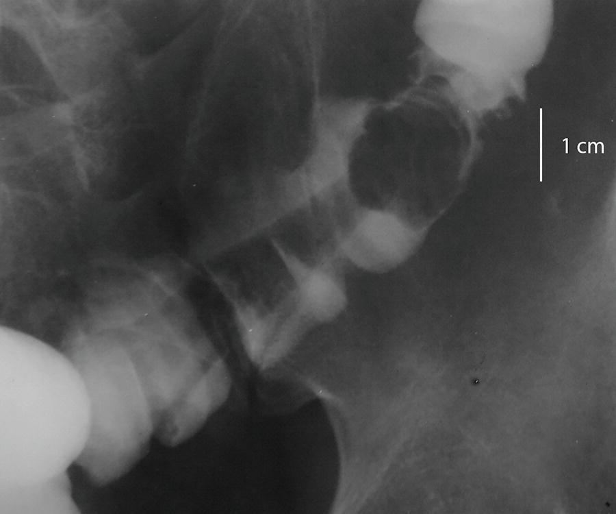
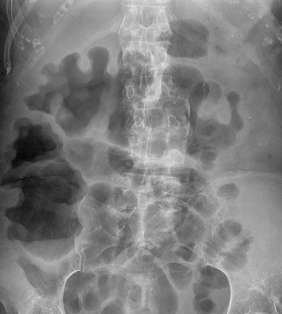
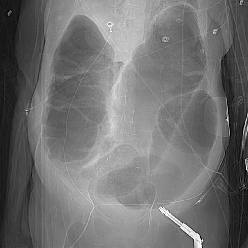
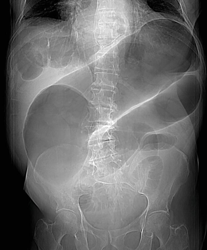
A. Repeat barium examination in 6 months to assess stability
B. Surgical consultation for sigmoid colectomy
C. Colonoscopy with snare polypectomy
D. Flexible sigmoidoscopy with biopsy
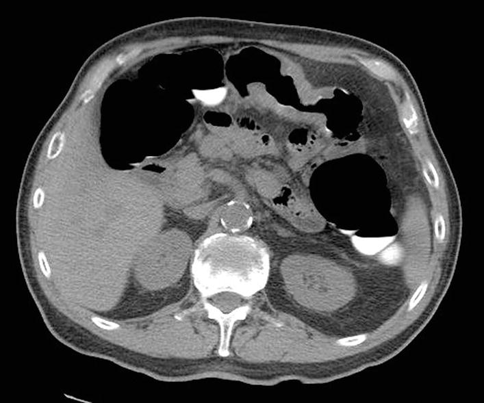
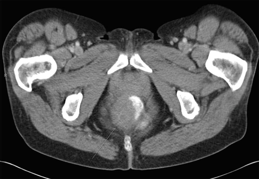
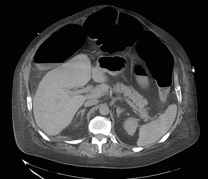
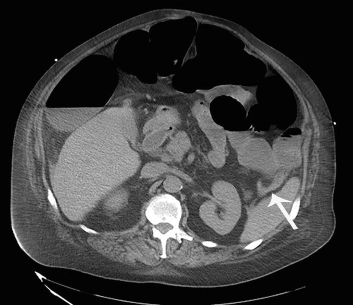
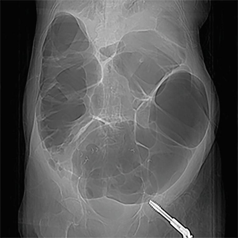
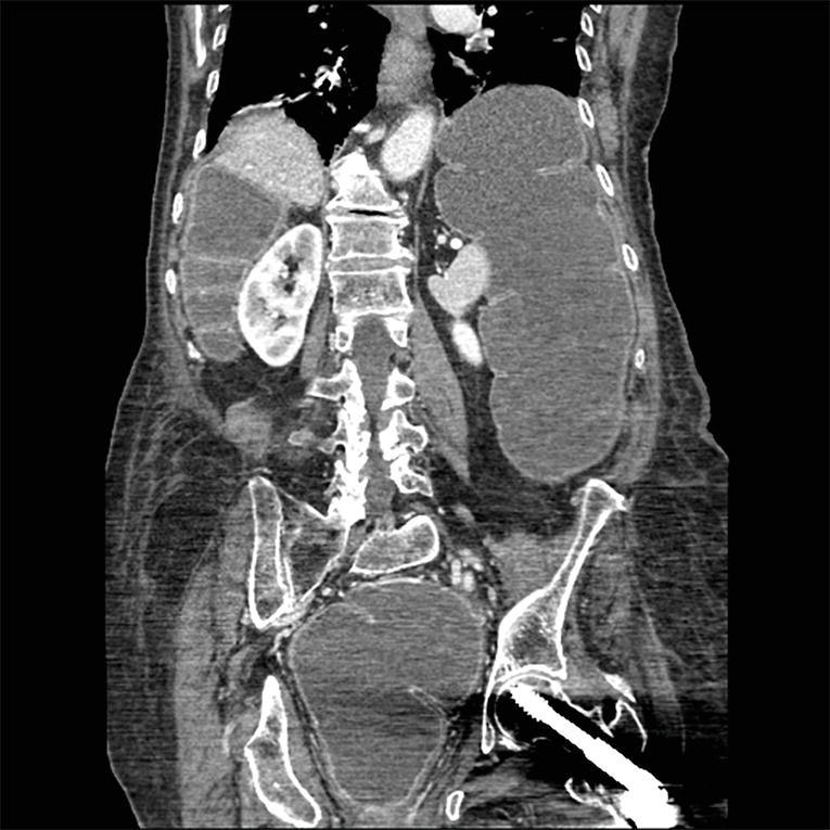
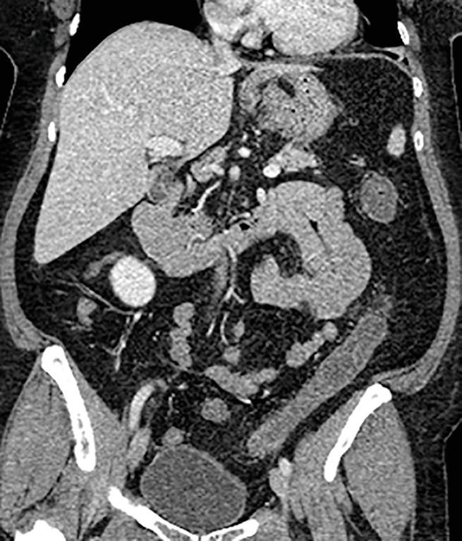
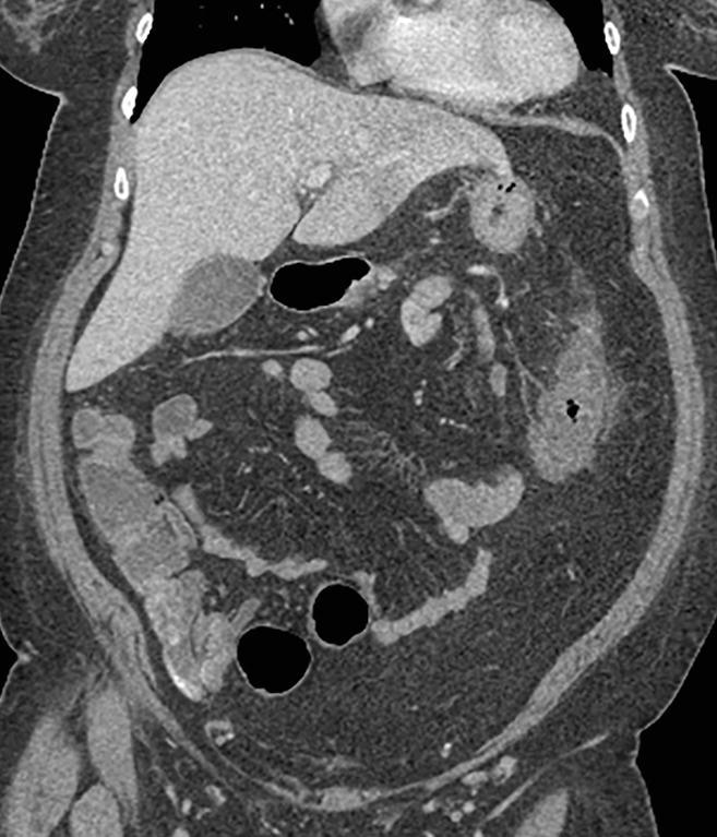
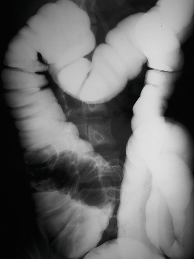
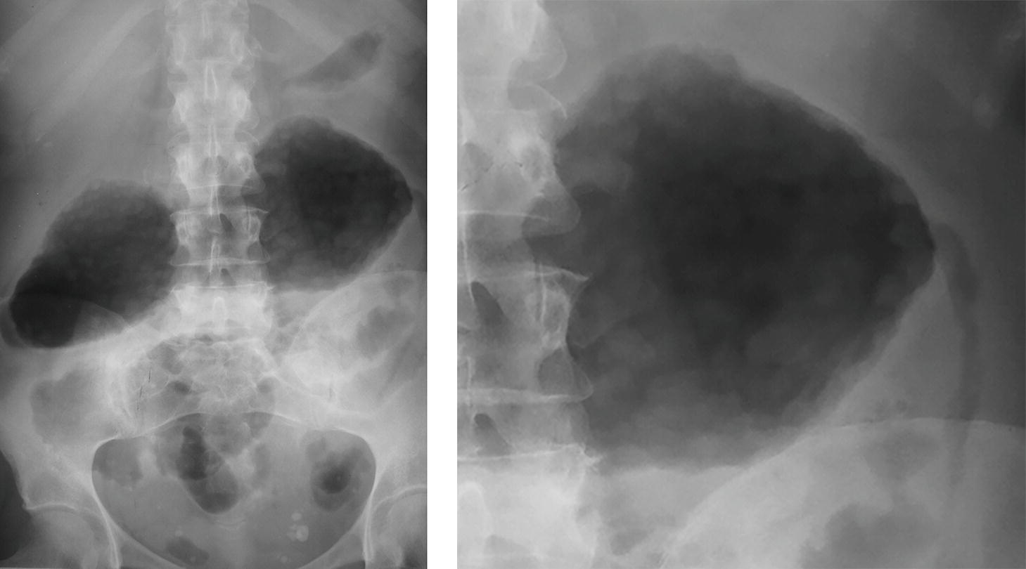
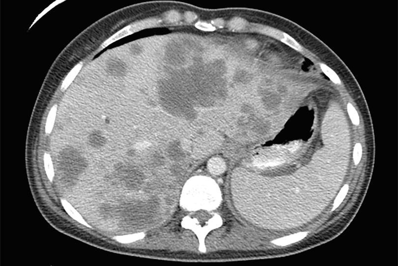
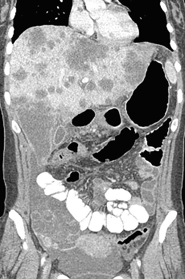
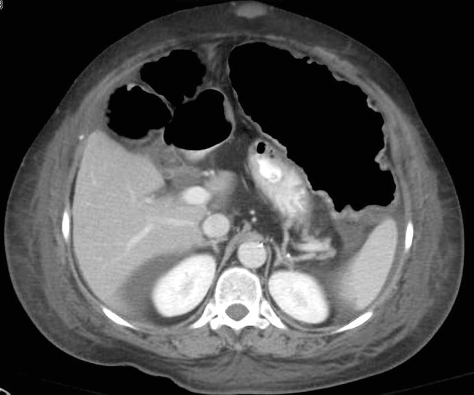
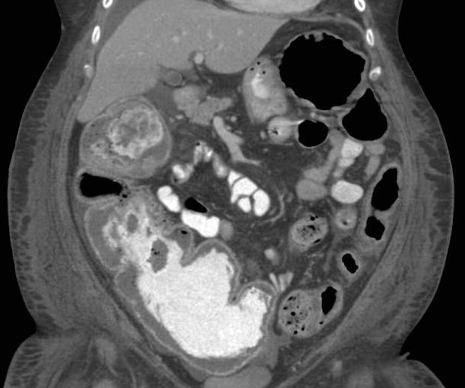
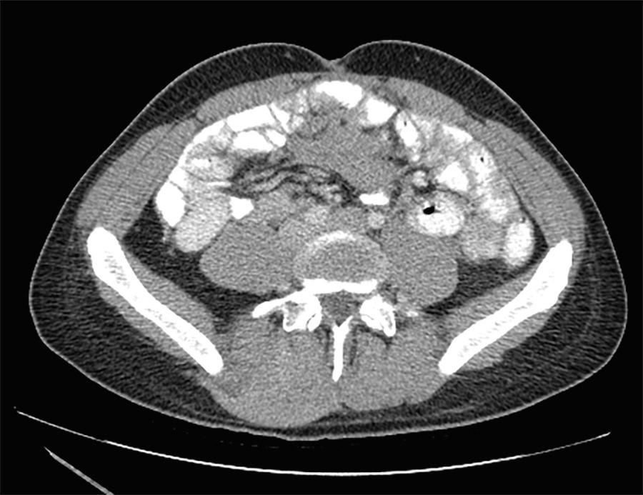
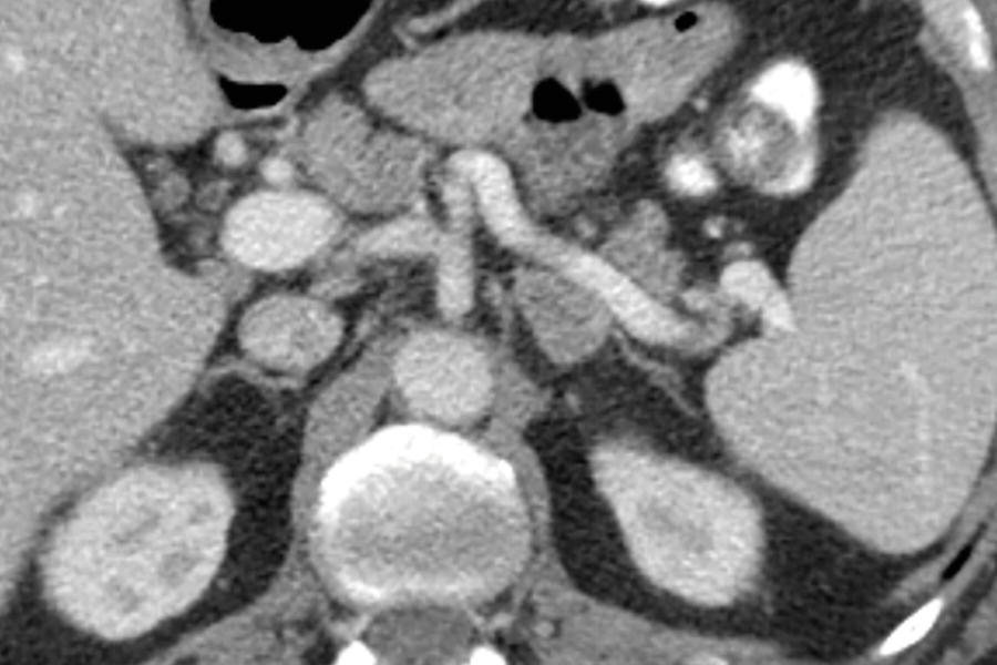
5 An 89-year-old male presents to the emergency room with abdominal pain, and a CT exam was performed. What is the most likely diagnosis?
A. Cecal volvulus
B. Cecal bascule
C. Sigmoid volvulus
D. Acute diverticulitis
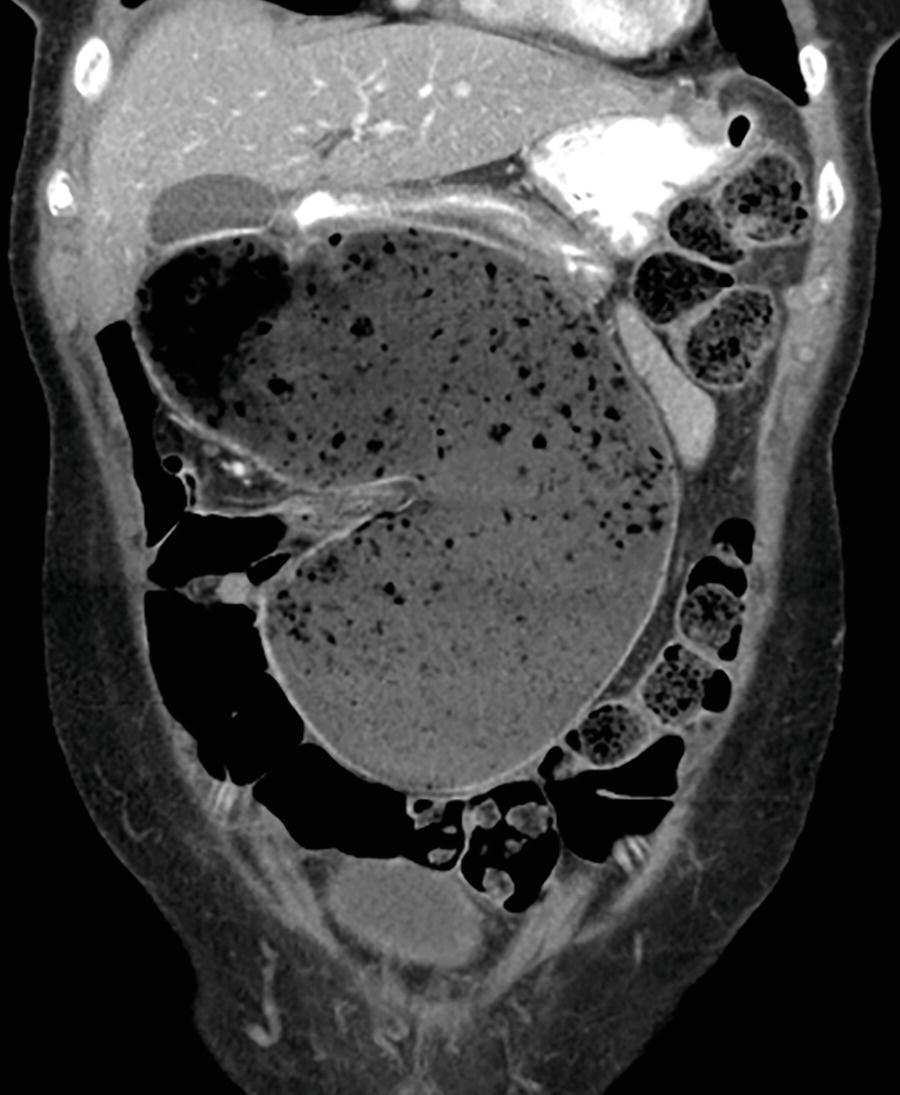
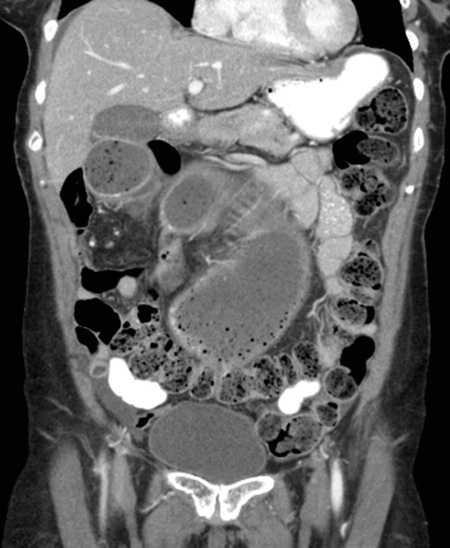
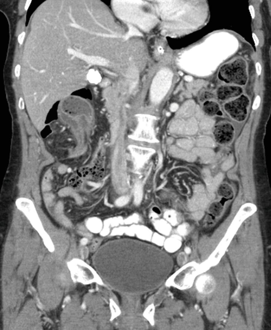
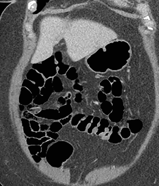
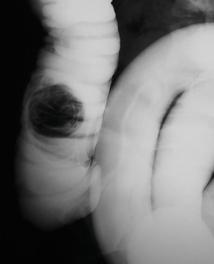
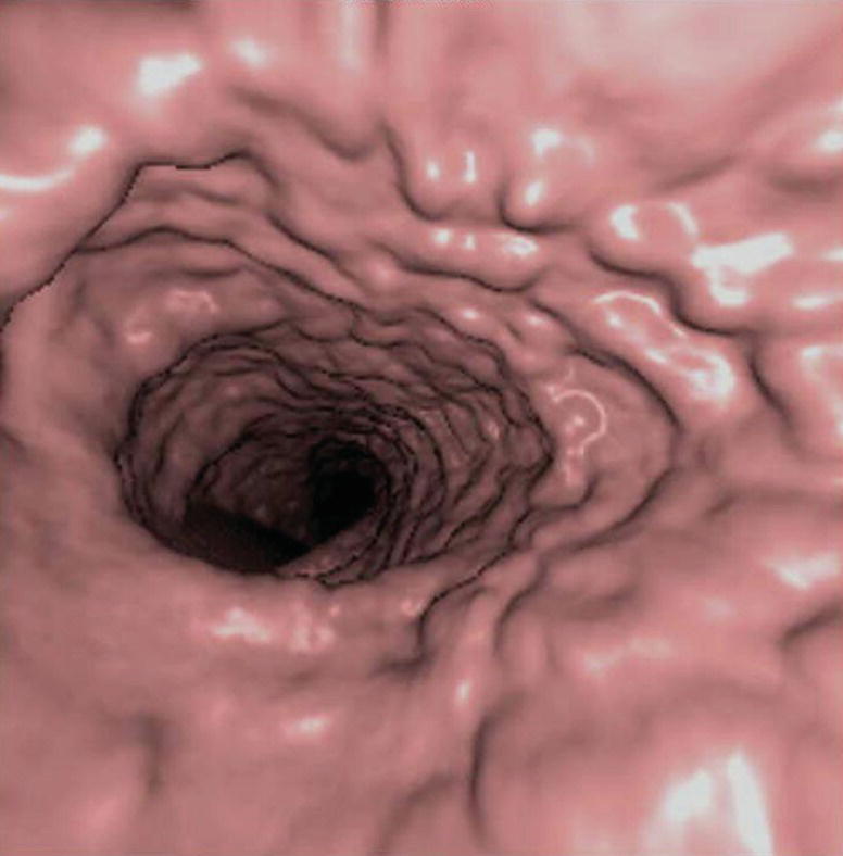
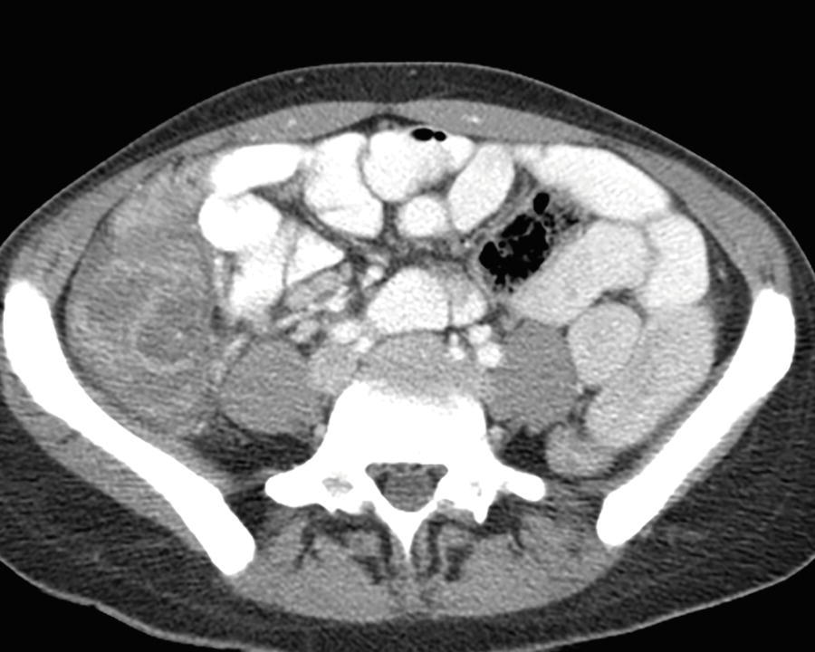
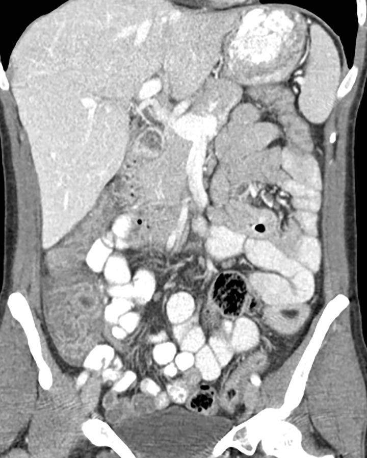
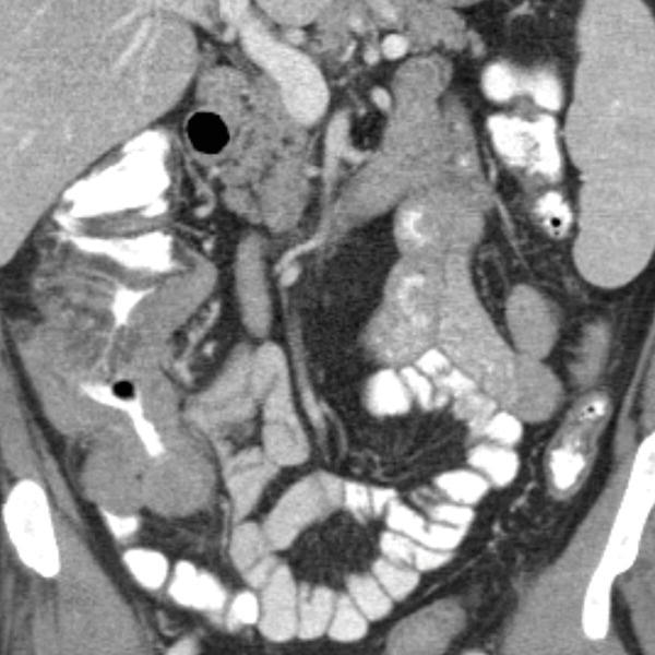
6 A 50-year-old female presents for her first screening CT colonography with the following finding. What is the most appropriate next step?
A. Surgical consultation to evaluate for bowel perforation
B. Surgical consultation to evaluate for bowel ischemia
C. Gastroenterology consultation to consider colonoscopy
D. If the patient is asymptomatic, she may be discharged with instructions to return if symptoms develop
7a The inpatient below presented with abdominal pain and diarrhea following a course of antibiotic therapy. The cause of this process is thought to be secondary to which of the following organisms?
A. Gram-negative anaerobe
B. Gram-positive anaerobe
C. Gram-negative aerobe
D. Gram-positive aerobe
7b The clinical manifestation of this process is thought to be caused by what toxin?
A. Toxin A
B. Toxin B
C. Toxin C
D. Toxin D
8 The risk of a 1.5-cm polyp harboring an invasive cancer is approximately
A. Less than 1%
B. Approximately 10%
C. Approximately 50%
D. Approximately 90%
9 A 70-year-old male presents for CT colonography following an incomplete colonoscopy. What is the most appropriate next step for this patient?
A. Routine screening colonoscopy in 10 years
B. Routine screening CT colonography in 5 years
C. Repeat colonoscopy now for biopsy if not already performed versus surgical consultation for resection
D. Capsule endoscopy to evaluate the proximal small and large bowel
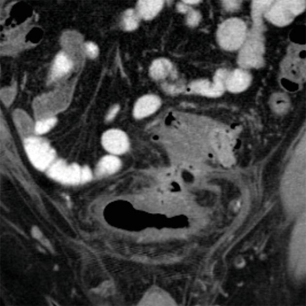
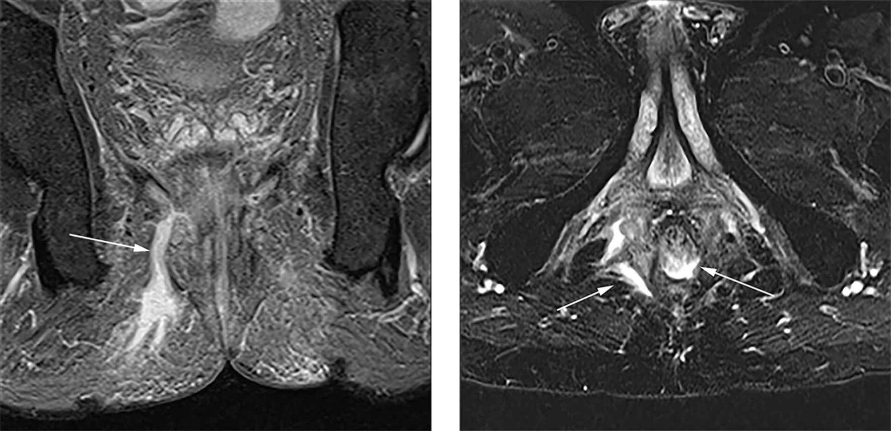
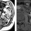
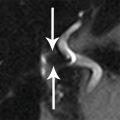
10 A 37-year-old man with known rectal adenocarcinoma presents for staging evaluation. An abdominopelvic MRI examination was performed. Based on the displayed image, what is the most likely T stage of disease?
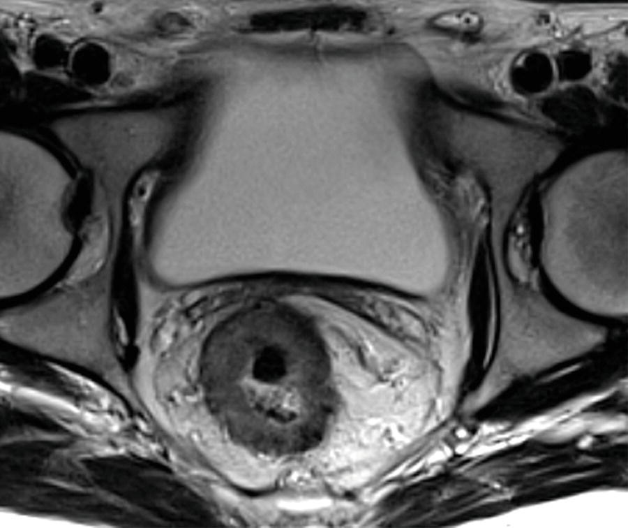
A. T1
B. T2
C. T3
D. T4
11 A 62-year-old female has isolated lung metastases from a colorectal neoplasm. Which of the following is the likely site of her primary tumor?
A.
B.
C.
D.
12 An 81-year-old male who has been in the intensive care unit for 2 weeks following a myocardial infarction develops abdominal bloating and diffuse abdominal pain. A CT obtained demonstrates a small bowel and colonic distention with a transition in caliber at the splenic flexure (arrow) without obvious wall thickening or extrinsic abnormality. The most likely diagnosis is
A. Pseudomembranous colitis
B. Acute colonic pseudoobstruction
C. Mechanical obstruction due to adhesions
D. Mechanical obstruction due to acute diverticulitis
13 In acute colonic pseudoobstruction, what is the most appropriate next step in management?
A. No change in management needed, as the condition is self-limited
B. Perform a single-contrast barium enema to exclude an occult neoplasm
C. Nasogastric decompression
D. Colonoscopic decompression
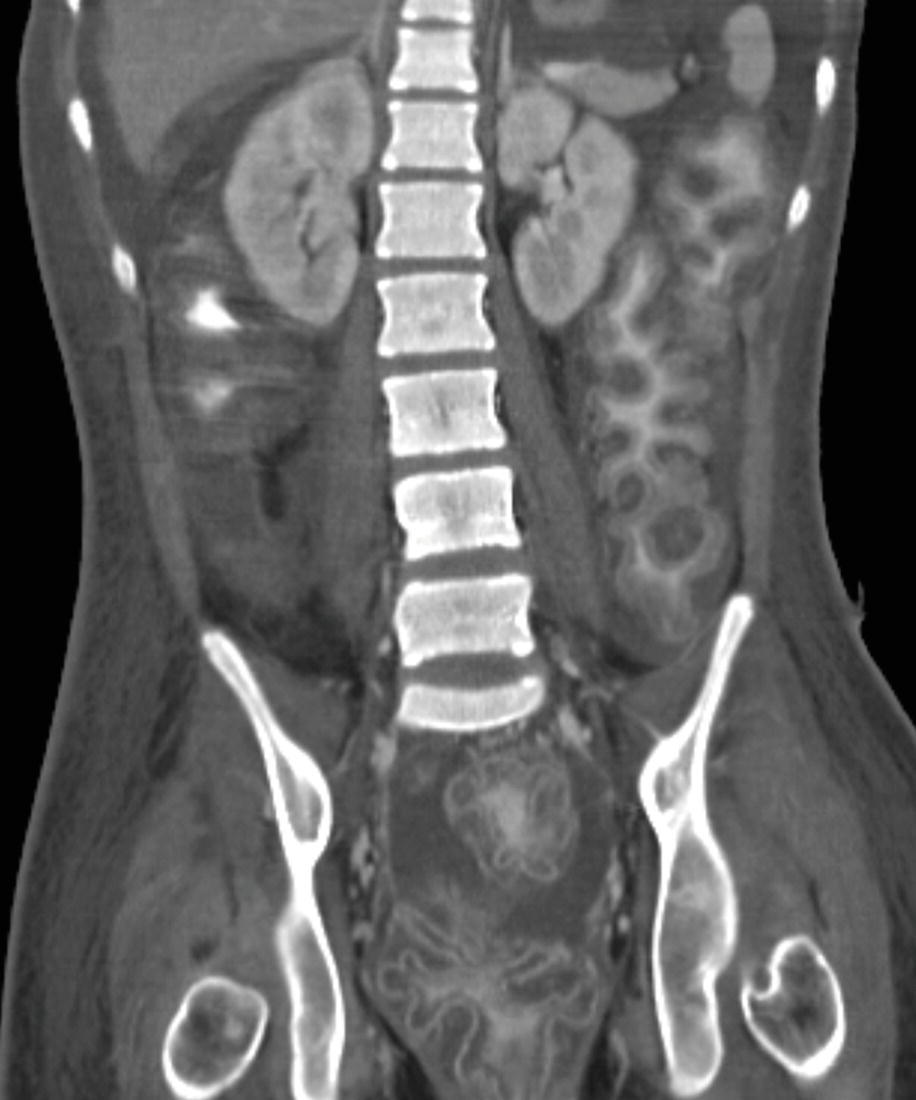
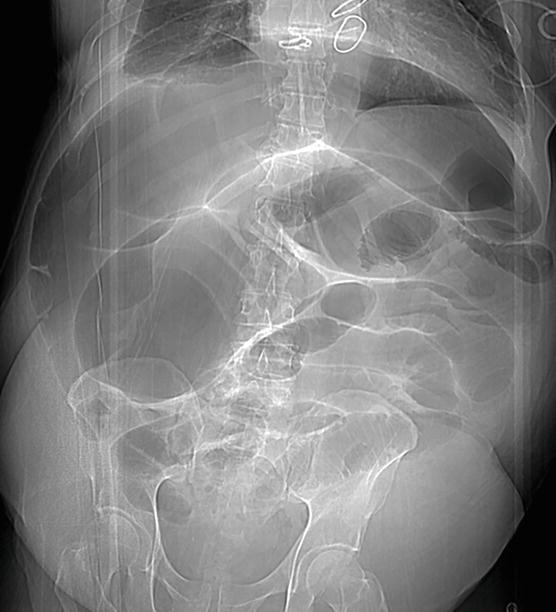
14 An 85-year-old female with chronic constipation is noted to have abdominal bloating and moderate abdominal discomfort. An abdominal CT is obtained.
No distal obstructing abnormality is found at colonoscopy and she is treated with rectal tube decompression. She has had multiple repeated admissions for the same presentation, including the scout from a CT 8 months earlier.
CT scout 8 months prior to the current admission.
A biopsy is performed of the bowel. What pathologic changes are most likely?
A. Fibrosis and amyloid deposition
B. Loss of intramural ganglion cells
C. Muscular hypertrophy
D. No pathologic abnormalities
15 An 88-year-old patient presents with abdominal pain and distention. What is the next most appropriate step regarding management of this patient?
A. Serial observation for a 24-hour period with discharge if hemodynamically stable
B. Emergent surgery
C. Endoscopic reduction of the torsion
D. Either B or C may be appropriate depending upon the clinical status of the patient
16 A 72-year-old female presents with pneumaturia. A CT scan is obtained. What is the most common cause of the findings below?
A. Colorectal cancer
B. Crohn disease
C. Diverticulitis
D. Pelvic radiation therapy
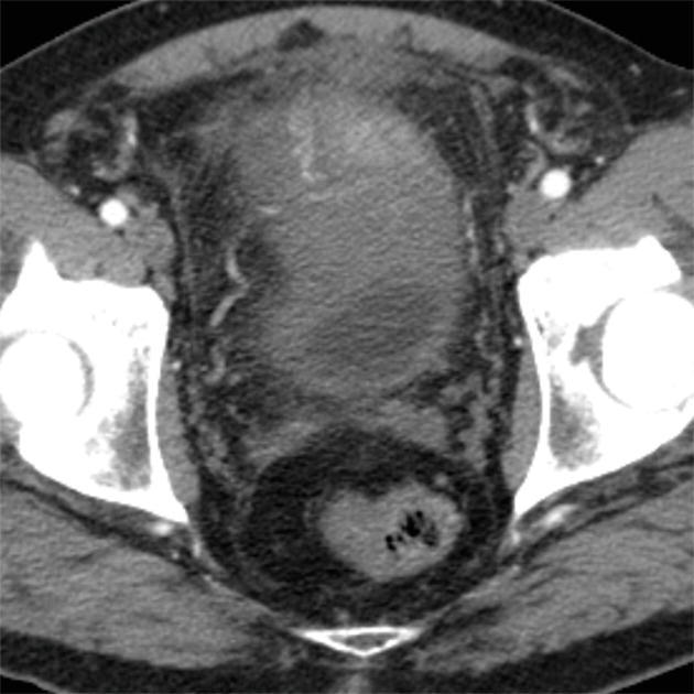
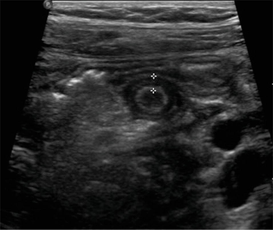
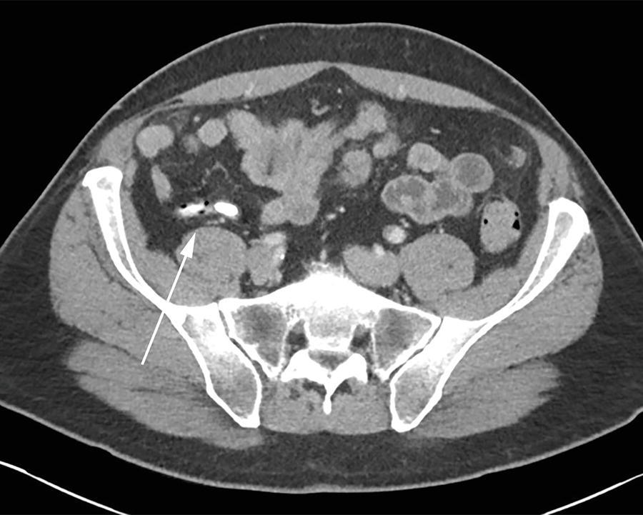
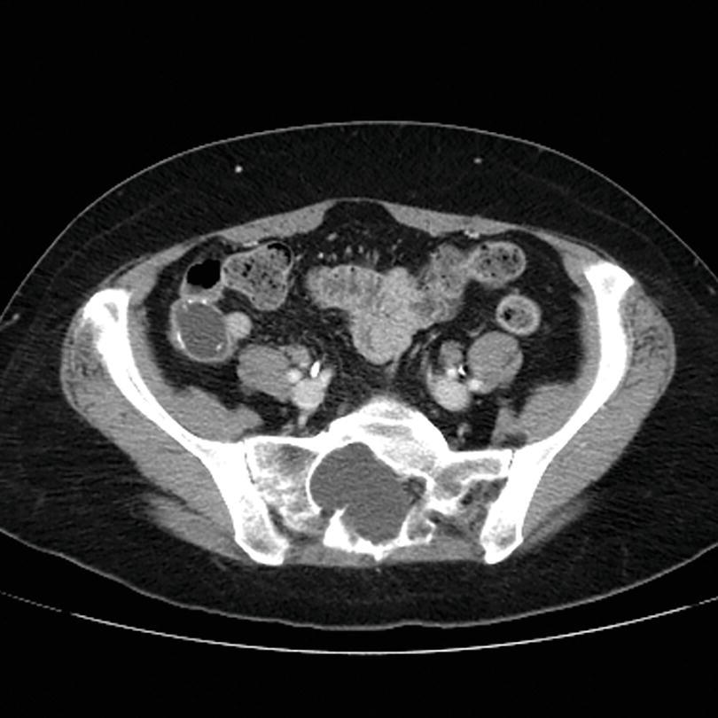
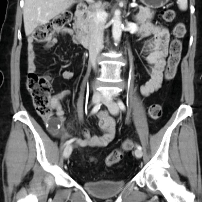
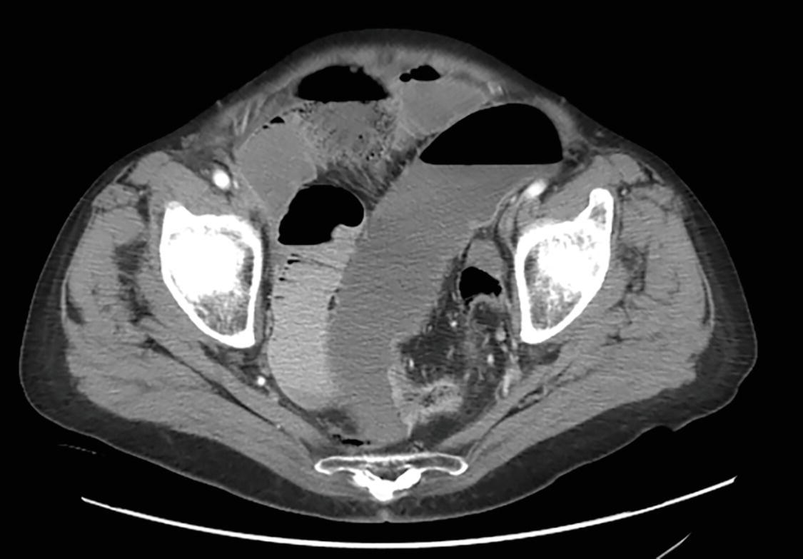
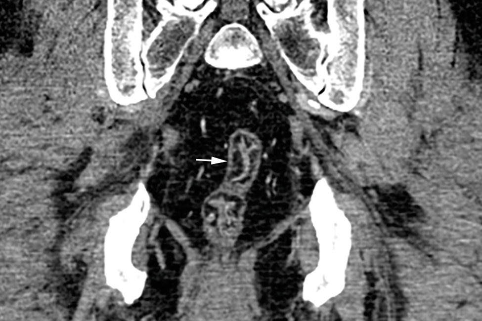
17a A 53-year-old patient presents with right lower quadrant pain. A CT is obtained. What is the likely diagnosis?
A. Sigmoid volvulus
B. Cecal volvulus
C. Acute appendicitis
D. Cecal bascule
17b What is the appropriate management for this patient?
A. Close observation for 24 hours and then discharge if stable
B. Exploratory laparotomy
C. Endoscopic decompression
D. Water-soluble enema reduction
18a A 35-year-old female patient presents to emergency room with right lower quadrant pain, nausea, and an elevated white blood cell count. The surgery team is concerned about acute appendicitis. She has a positive pregnancy test. What is next best imaging test for this patient?
A. Low-dose CT without intravenous contrast
B. MRI of the abdomen and pelvis without intravenous contrast
C. Ultrasound of the pelvis, targeted to the right lower quadrant
D. Indium-111 white blood cell scan
18b Ultrasound of the pelvis was performed. Which of the following is TRUE regarding the role of ultrasound imaging in patients with suspected appendicitis?
A. Noncompressible blind-ending tubular structure with a diameter >6 mm at the point of maximum tenderness can be considered diagnostic for appendicitis.
B. Presence of periappendiceal fluid on ultrasound exam suggests a diagnosis of ruptured appendicitis.
C. The normal peristalsing small bowel loops in right lower quadrant virtually excludes appendicitis, if the appendix is not visualized.
D. The nonvisualization rate (for a normal appendix) is 10% to 20% on ultrasound.
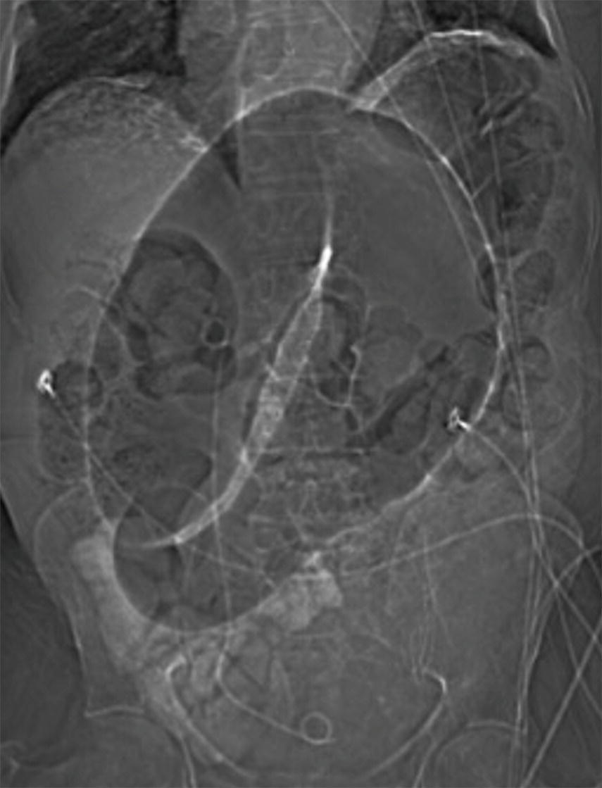
19 An 85-year-old male presents with intermittent bright red blood per rectum and a hematocrit of 28. He receives two units of packed red blood cells. Initial colonoscopy fails to detect the site of bleeding. At 11 pm, he becomes hypotensive, develops an episode of ongoing hemorrhage, and has the CT visceral angiogram study below. Following the CT, the bleeding stops, and his blood pressure stabilizes. What is the next step in management?
A. Send directly to the angiography suite for catheter embolization.
B. Schedule a repeat emergent colonoscopy within 2 hours.
C. Schedule sigmoid resection within 24 hours.
D. Repeat study with oral contrast to look for an underlying colonic neoplasm.
E. Provide supportive care and observation with reassessment in the morning.
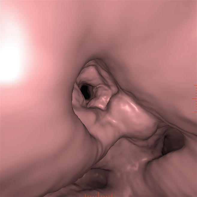
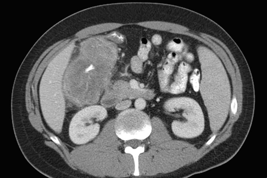
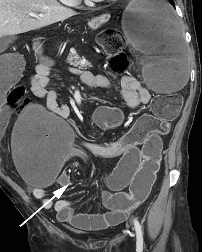
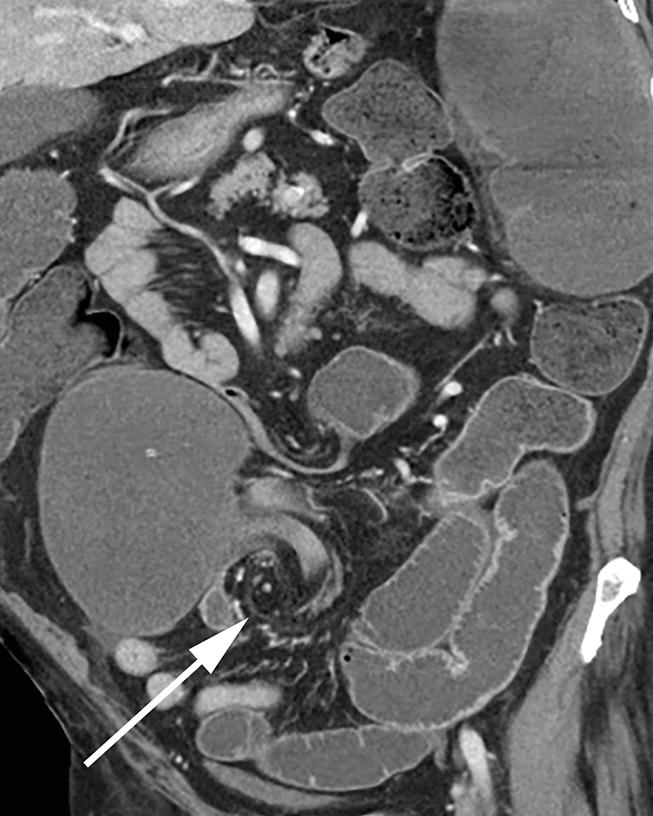
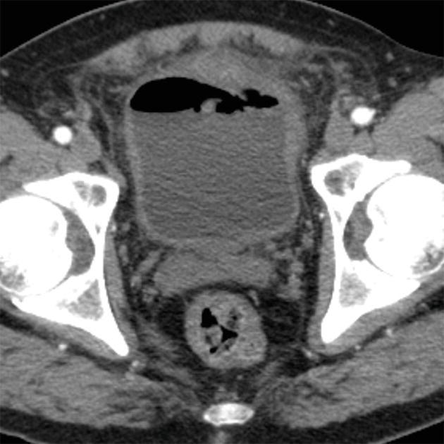
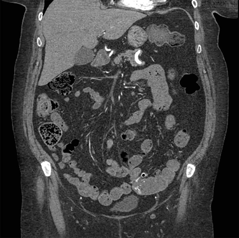
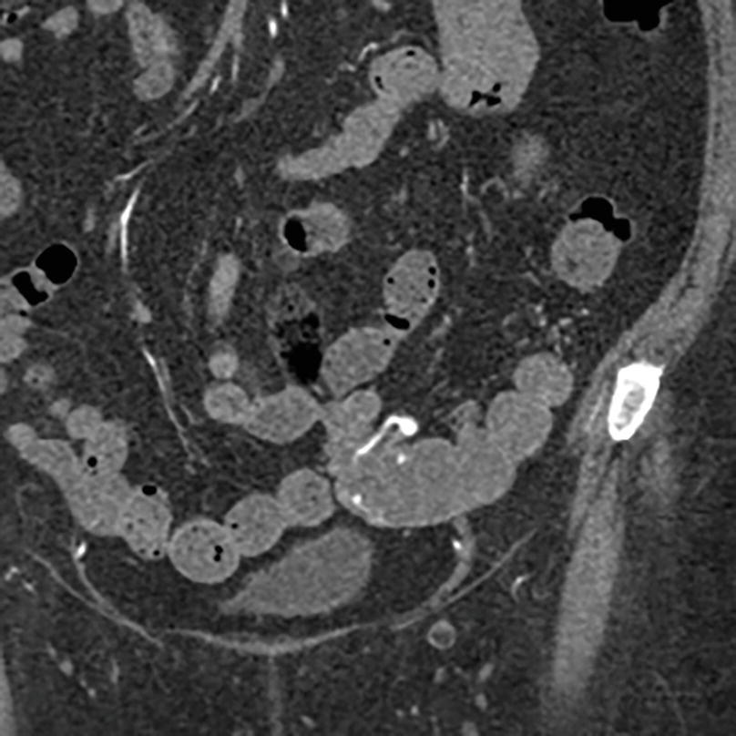
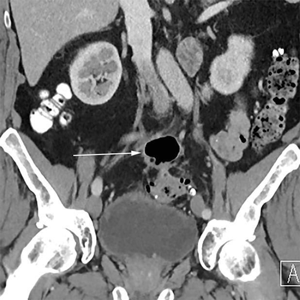
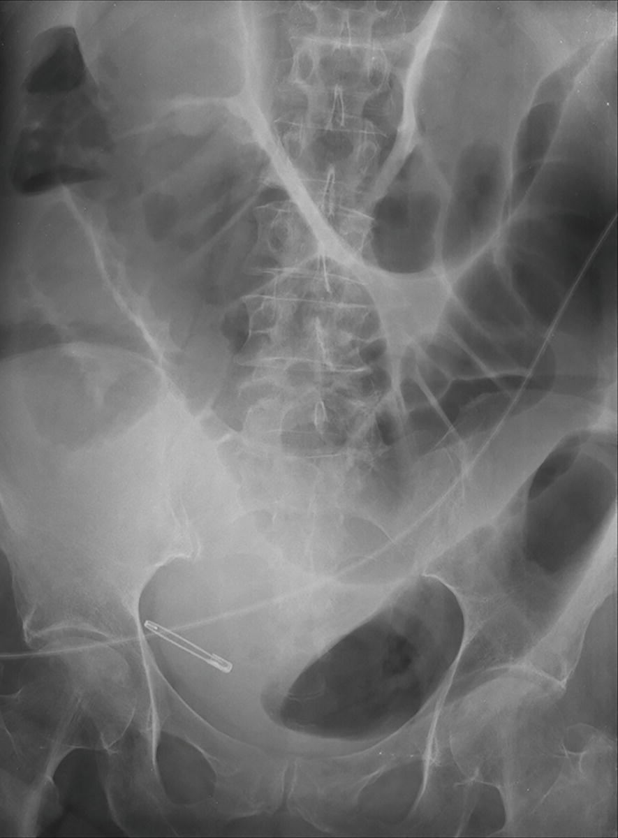
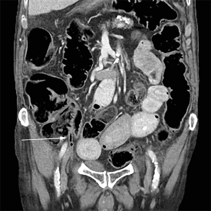
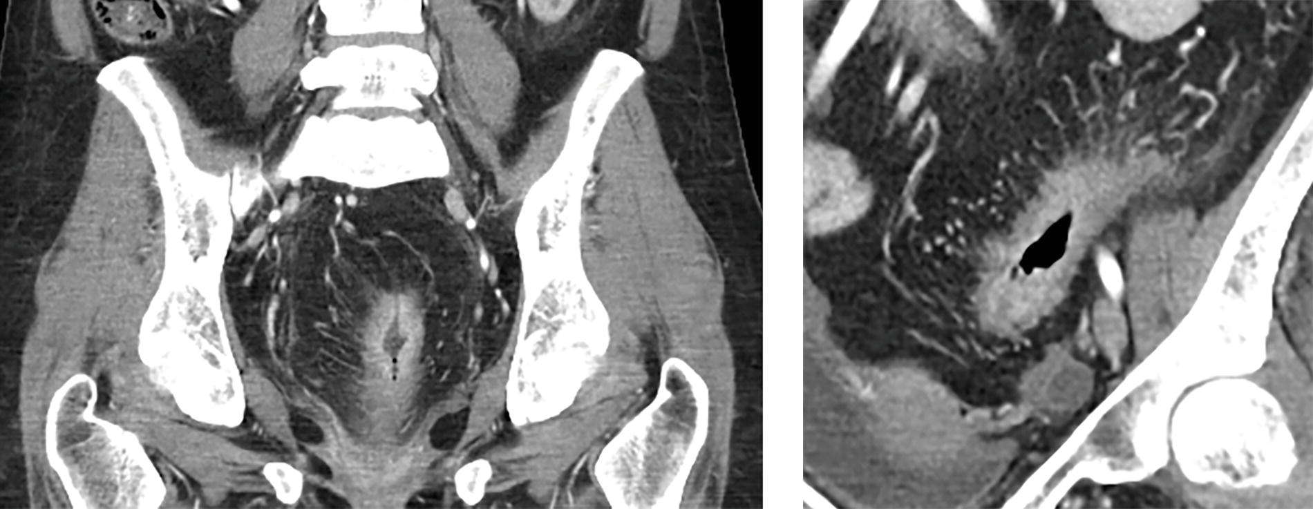
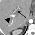
20 This 45-year-old male patient has a CT for staging of a testicular cancer. He has never received enteric contrast. The patient is asymptomatic with no fever or leukocytosis. The structure below is connected to the cecal tip. What would be the best follow-up?
A. No imaging follow-up necessary
B. 6-month low-dose CT follow-up to ensure resolution of the findings
C. Ultrasound to assess for acoustical shadowing
D. Indium-111 WBC scan
21a A 45-year-old male presents with acute-onset left lower abdominal pain of 2 days duration. He presents to the emergency department and is found to be afebrile with a normal WBC count. A CT of the abdomen and pelvis is obtained. What is the most likely diagnosis?
A. Acute appendicitis
B. Diverticulitis
C. Omental infarct
D. Epiploic appendagitis
21b The most appropriate management for the patient would be
A. Analgesics and outpatient observation
B. Emergent surgical consultation
C. Inpatient admission and intravenous antibiotics
D. Oral antibiotics
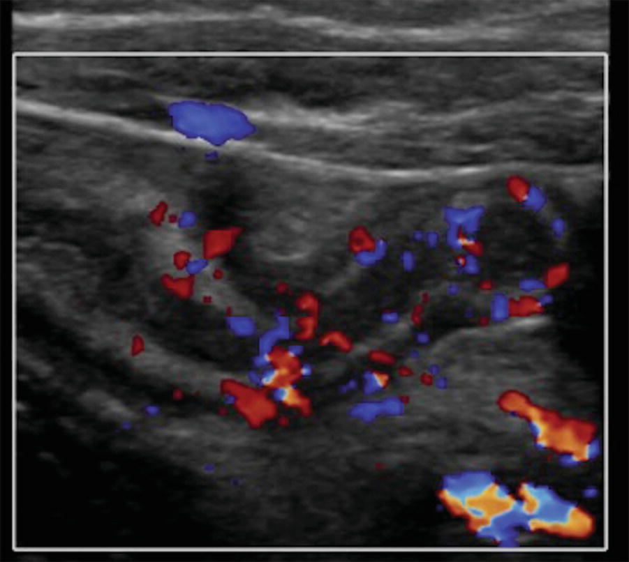
22a A 55-year-old female presents with mild right lower quadrant tenderness. A CT scan was performed. What is the cause of the pertinent finding?
A. Acute appendicitis
B. Tarlov cyst
C. Appendiceal cystadenoma
D. Meckel diverticulitis
22b What is the most appropriate next step in management?
A. Surgical consultation for laparoscopic appendectomy
B. Surgical consultation for right hemicolectomy
C. Oncologic consultation for chemotherapy
D. Follow-up CT in 1 year
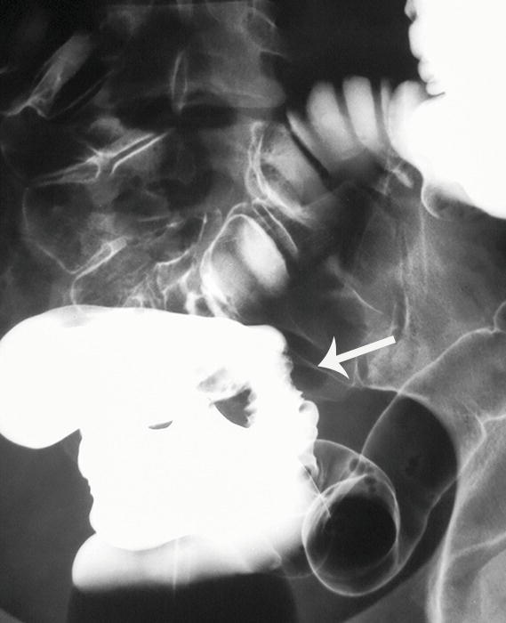
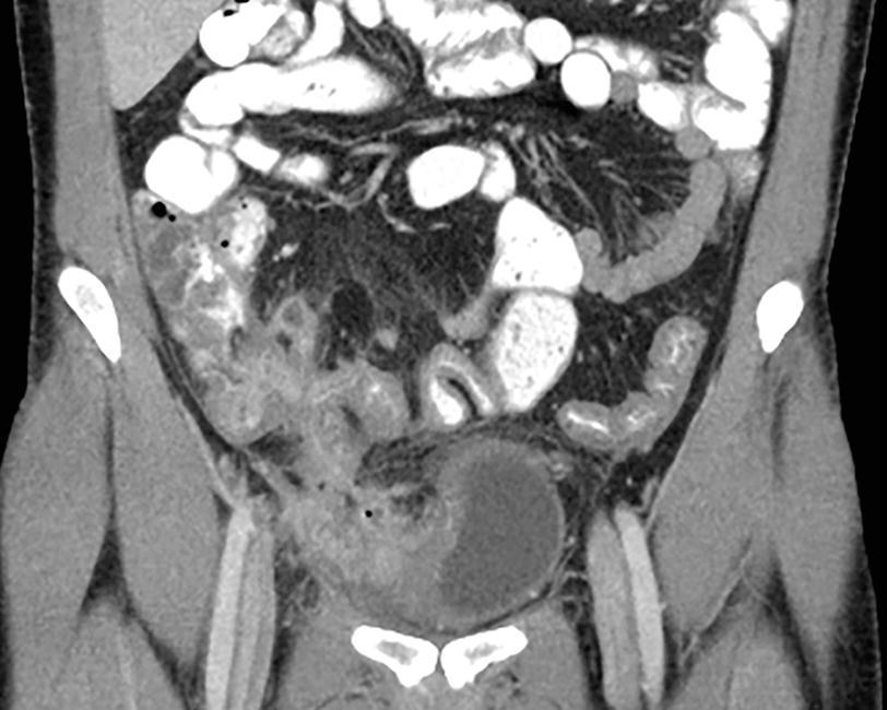
23 A 65-year-old male with a history of previous episodes of diverticulitis presents with fever, mild leukocytosis, and lower abdominal pain. What does the finding below on the CT most likely represent?
A. Colovesical fistula
B. Communicating duplication cyst
C. Cecal bascule
D. Giant inflammatory sigmoid diverticulum
24 An 89-year-old female presents with acute-onset abdominal pain and hematochezia. Which clinical scenario best fits the finding?
A. History of antibiotic therapy for pneumonia
B. History of cardioversion for cardiac arrhythmia
C. Recent overseas travel
D. History of prior episodes of diverticulitis
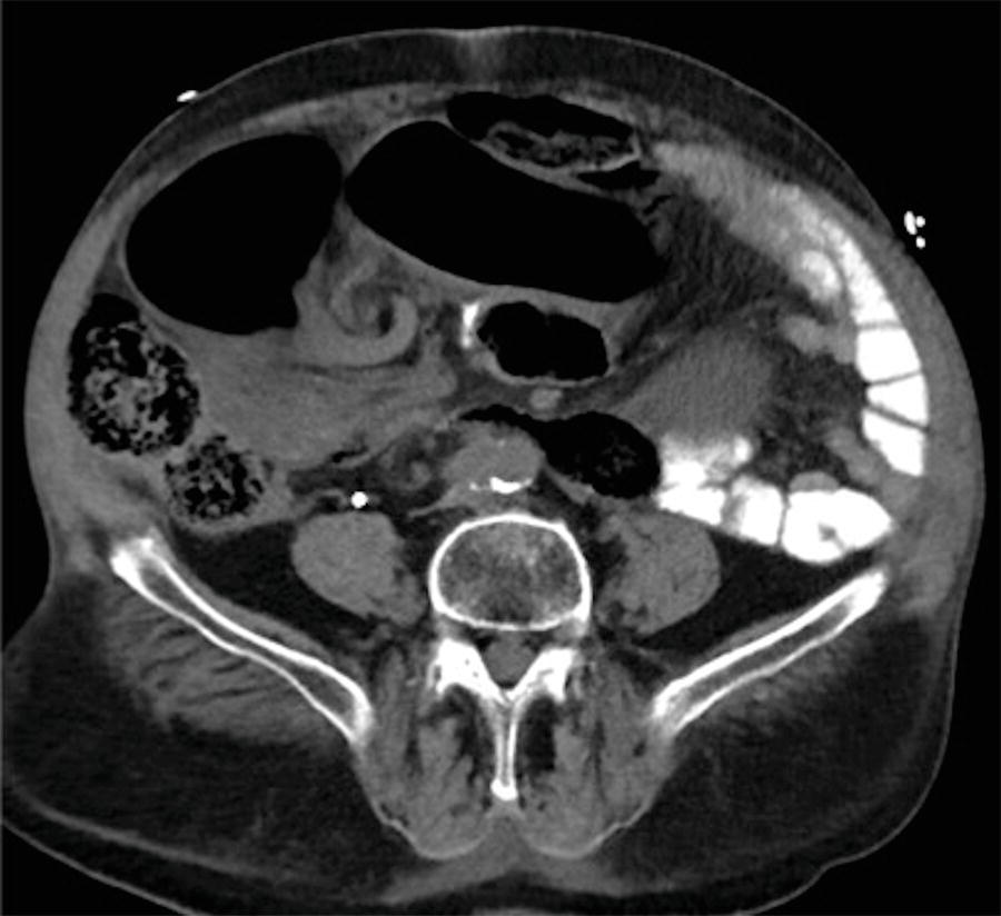
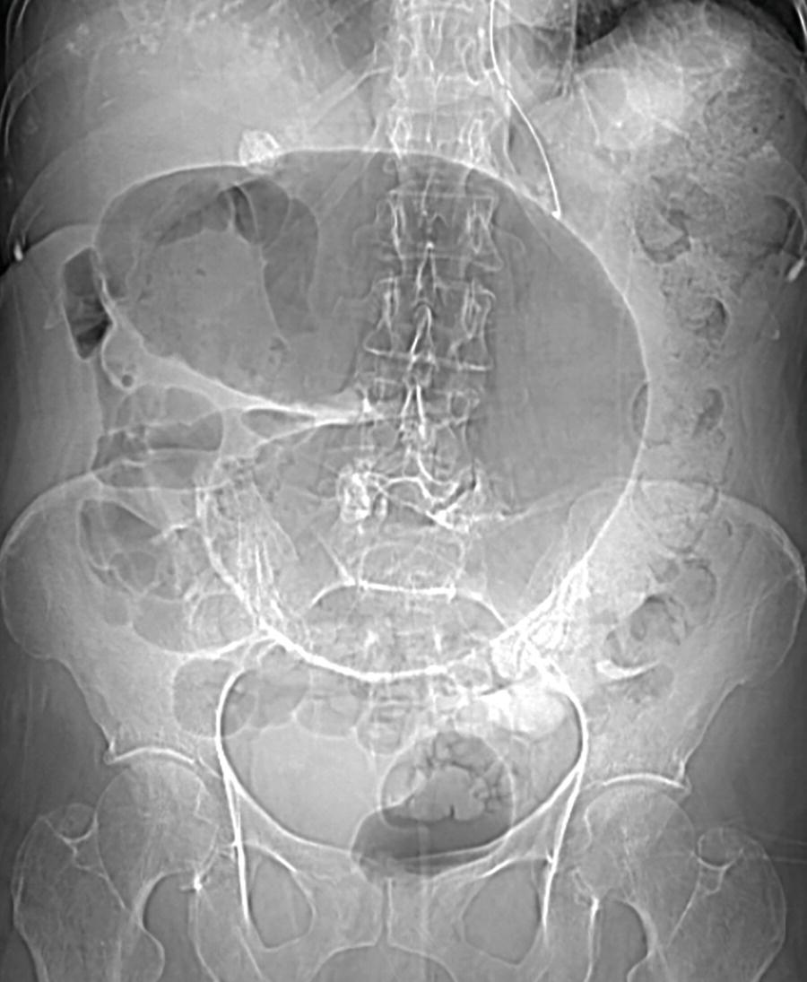
25 An 86-year-old male presents with chronic abdominal pain, constipation, and bloating. An abdominal radiograph and a CT are obtained. Approximately what percentage of large bowel obstructions does this finding account for?
A. 95% or greater
B. 60%
C. 20%
D. Less than 5%
26 The most common site for perforation due to an obstruction from a sigmoid colonic neoplasm is at the
A. Cecum
B. Splenic flexure
C. Sigmoid just proximal to the obstruction
D. At the level of the neoplasm
27 Match the following type of infection with the typical distribution of the colitis. Each answer may be used once, more than once, or not at all.
A. Salmonella typhi
B. Mycobacterium tuberculosis
C. Chlamydia trachomatis (lymphogranuloma venereum)
D. Entamoeba histolytica
E. Shigella
F. cytomegalovirus
G. E. coli
1. More often right-sided
2. More often left-sided
3. Most commonly diffuse
28a A 36-year-old female has a history of intermittent abdominal pain. She undergoes a barium enema. Full abdominal film and spot views of the sigmoid are performed. What are your interpretations of the findings?
A. Normal examination
B. Finding suspicious for a colonic neoplasm
C. Acute ulcerative colitis changes
D. Diverticulitis
28b The patient in this case undergoes a colonoscopy, and while luminal narrowing is encountered in the sigmoid, no mucosal abnormality is seen. What is the next best step?
A. Surgical exploration
B. CT colonography
C. MR pelvis with T1-weighted fat-suppressed imaging
D. Pelvic ultrasound
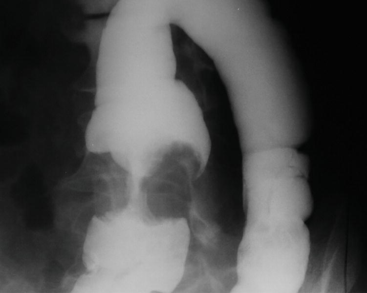
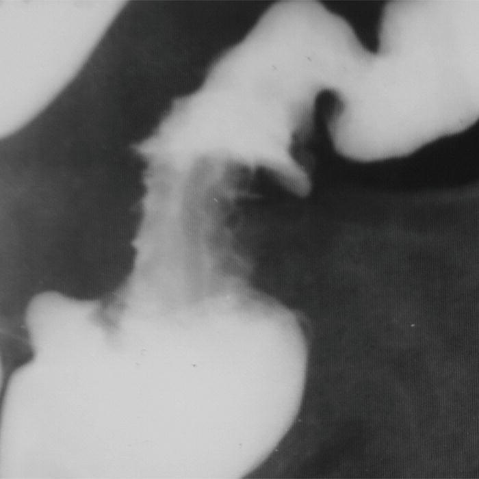
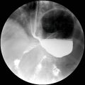
29a A 25-year-old patient presents with acute abdominal pain. Plain films suggest a small bowel obstruction, and the patient undergoes a single-contrast barium enema. What is the cause of the finding?
A. Lipomatous ileocecal valve
B. Ileocolic intussusception
C. Appendiceal mucocele
D. Inverted appendiceal stump
29b Regarding the finding above, what are likely associated findings in this 25-year-old patient?
A. History of perianal fistulae
B. Mucocutaneous pigmentation perioral region
C. Gluten sensitivity
D. Gastroesophageal reflux disease and sclerodactyly
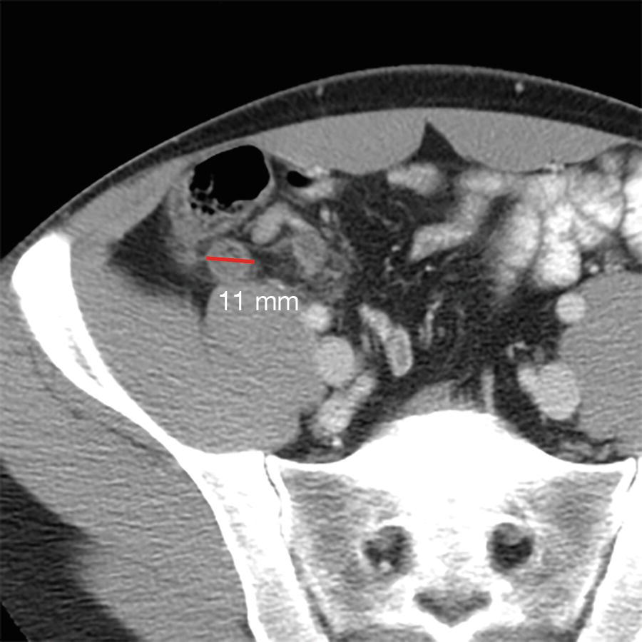
30 A 79-year-old male has a history of mild abdominal pain and right groin fullness. He has a CT of the abdomen and pelvis. What is the name of the finding?
A. Littre hernia
B. Amyand hernia
C. Richter hernia
D. De Garengeot hernia
31 A 56-year-old female undergoing chemotherapy for acute myelogenous leukemia presents with abdominal pain and watery diarrhea. The patient is afebrile and has a WBC count of 850 cells/mm3. She undergoes a CT scan. Regarding neutropenic colitis, which of the following statements is TRUE?
A. The finding more commonly involves the left colon.
B. Pneumatosis intestinalis is an indication for emergent surgery.
C. The radiographic appearance is distinct from that of C. difficile colitis.
D. The cause is likely multifactorial with ischemia, hemorrhage, and infection among possible contributing factors.
32 In the patients below with inflammatory bowel disease, match the image with the diagnosis with the strongest association. Each answer may be used once, more than once, or not at all.
A. Acute ulcerative colitis
B. Active Crohn enterocolitis
C. Either disease in chronic phase
D. Neither disease
1. 39-year-old female
2. 27-year-old male
3. 20-year-old male
4. 45-year-old female
5. 18-year-old male
6. 52-year-old male
7. 32-year-old male
8. 28-year-old male
9. 48-year-old female
33 A 28-year-old patient recently completed a course of oral and topical treatment for ulcerative colitis and is currently asymptomatic. What term best describes the polyps seen in the double-contrast barium enema?
A. Adenomatous polyps
B. Acute inflammatory polyps
C. Postinflammatory polyps
D. Pseudopolyps with a background of submucosal ulceration
34a A 29-year-old female presents with abdominal pain and fullness. The following CT images are obtained. A colonoscopy is performed and biopsy of the cecal mass reveals adenocarcinoma. The patient’s father died at age 48 with colon cancer, and a maternal aunt was recently diagnosed with a large colonic polyp. What is the most likely diagnosis?
A. Familial adenomatosis polyposis
B. Hereditary nonpolyposis colorectal cancer (Lynch syndrome)
C. Gardner syndrome
D. Cronkhite-Canada syndrome
34b What additional imaging should be considered for this patient?
A. PET scan
B. Octreotide scan
C. Pelvic ultrasound and endometrial sampling
D. Thyroid ultrasound
35 A 35-year-old female with abdominal pain and distention, fever, and bloody diarrhea undergoes a contrast-enhanced CT. What is the most likely diagnosis?
A. Toxic megacolon
B. Familial adenomatous polyposis
C. Crohn disease
D. Lymphoid hyperplasia
36 A 20-year-old male presents with a palpable abnormality at the angle of the jaw.
What additional study should be considered?
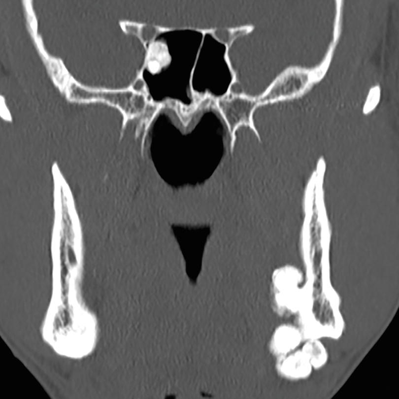
A. Double-contrast barium enema or colonoscopy
B. CT enterography
C. MRCP
D. Brain MRI with contrast
37 A 65-year-old male presents for evaluation of a possible lung nodule on a CXR. A chest CT is performed as an outpatient, and the following CT image is obtained in the upper abdomen. The patient is detained by the CT technologist who checks the images with the radiologist. The patient is anxious to leave for a lunch appointment. What is the next best step?
A. No abnormality is seen, and no further investigation is needed.
B. Pneumatosis coli is present, but the finding is interpreted as benign given the patient is asymptomatic.
C. The patient should be directed to the emergency department with a surgical consultation requested.
D. Additional image manipulation should be performed before making a decision as to the patient disposition.
38 A 35-year-old patient presents with the following findings:
Which of the following genetic abnormality is present?
A. Germ-line mutation in APC gene on chromosome 5q21
B. Defect in a DNA mismatch repair gene
C. Mutation in the PTEN gene on arm 10q
D. A germ-line mutation of the STK11/LKB1 tumor suppressor gene
39 A 25-year-old female presents with abdominal pain, fever, and bloody diarrhea of 4 days onset. She has no history of prior similar episodes and is otherwise healthy. The emergency department physician is concerned for acute appendicitis and orders a CT of the abdomen and pelvis. What do you recommend next?
A. Surgical consultation for acute appendicitis
B. Intravenous corticosteroids for acute ulcerative colitis flare
C. Stool cultures and empiric antibiotics
D. CT angiogram for suspected embolic ischemic colitis
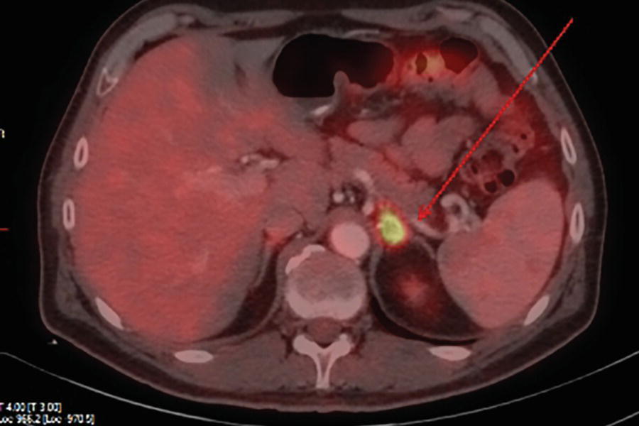
40 A 27-year-old HIV-positive patient presents with abdominal pain. A CT followed by a PET-CT is performed. What is the most likely diagnosis?
Related posts:
Stay updated, free articles. Join our Telegram channel

Full access? Get Clinical Tree


