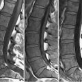
43 Compression Fractures The typical MRI appearance of an acute, osteoporotic compression fracture is illustrated in Figs. 43.1A,B. On (A) sagittal FS T2WI, high SI edema is present throughout the vertebral body, while the normal high vertebral SI on (B) T1WI is replaced by low SI edema. Superior end plate compression deformity and resultant height loss is also present. Figures 43.2A through 43.2D illustrate a less common appearance of such fractures. Here, the T12 vertebral body demonstrates height loss, low SI within the marrow due to edema, and a pocket of low SI fluid on (A) T1WI. Due to the use of FSE technique, vertebral body SI appears almost normal in the T2WI of Fig. 43.2B, although the fluid pocket is better visualized. On (C) FS T2WI the fluid pocket is again well seen, together with the edema within the adjacent marrow. (D) Contrast-enhanced FS T1WI show the fluid pocket as low SI surrounded by avidly enhancing tissue, the latter likely correlating with damaged, leaky capillaries resulting from the fracture present. Chronic, benign fractures lack edema and may be somewhat subtle (reflected by height loss alone). The L5 vertebral body in this case had demonstrated a loss of height, from end plate to end plate, since the prior examination, and exhibits reduced height versus the other vertebrae in Fig. 43.2
![]()
Stay updated, free articles. Join our Telegram channel

Full access? Get Clinical Tree


