Congenital Pulmonary Airway Malformation
Melissa L. Rosado-de-Christenson, MD, FACR
Key Facts
Terminology
Abnormal mass of lung tissue with varying degrees of cystic change
Imaging Findings
Unilateral multilocular thin-walled cysts
Air-filled, fluid-filled, or air-fluid levels
Dominant cyst surrounded by smaller cysts
Rarely solid mass
Mass effect on adjacent structures
Associated congenital anomalies
Antenatal diagnosis with ultrasound or MR
Top Differential Diagnoses
Congenital Lobar Overinflation
Pulmonary Sequestration
Bronchogenic Cyst
Neuroblastoma
Pathology
Classification based on lesion components reflecting morphology of portions of tracheobronchial tree
Type 1 lesions most frequently diagnosed
Clinical Issues
Neonate with progressive respiratory distress
Antenatal diagnosis with ultrasound
Difficult confident histologic diagnosis in older children and adults due to superimposed acute and chronic inflammation
Treatment with surgical excision
Good prognosis for type 1 lesions
Diagnostic Checklist
Unilateral cystic or solid pulmonary mass in infancy
Characterization with prenatal ultrasound or MR
Postnatal evaluation with chest CT in selected cases
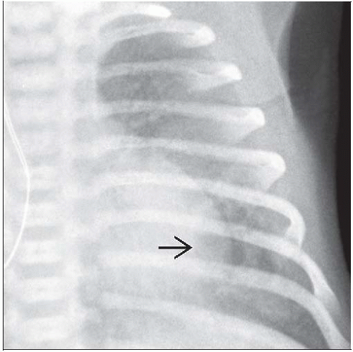 Frontal radiograph coned-down to the left lung shows a focal left lower lobe air-filled multicystic type 1 congenital pulmonary airway malformation. A dominant air-filled cyst  is surrounded by smaller air-filled cysts. is surrounded by smaller air-filled cysts. |
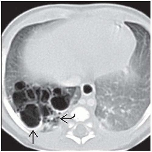 Axial CECT shows a right lower lobe multicystic type 1 CPAM. Larger air-filled cysts  are surrounded by smaller cysts are surrounded by smaller cysts  . The cyst walls are relatively thin without nodular components. . The cyst walls are relatively thin without nodular components. |
TERMINOLOGY
Definitions
Congenital pulmonary airway malformation (CPAM)
Currently accepted terminology
Spectrum of lesions affecting various portions of tracheobronchial tree and distal airways
Congenital cystic adenomatoid malformation (CCAM)
Outdated terminology
Some lesions are not cystic
Most lesions are not adenomatoid
CPAM: Abnormal mass of pulmonary tissue with varying degrees of cystic change
Adenomatoid: Term describes intralesional appearance of gland-like back-to-back airways
Intralesional bronchioles, alveolar ducts, and alveoli
Airways lined by cuboidal epithelium
Cystic: Term describes intralesional appearance of air-or fluid-filled bronchial-like or bronchiolar-like spaces
CPAM: Communicates with tracheobronchial tree; typically normal blood supply and venous drainage
IMAGING FINDINGS
General Features
Best diagnostic clue
Multilocular cystic pulmonary lesion in fetus, neonate, or infant
Rarely diagnosed beyond infancy as chronic inflammation may preclude confident histologic diagnosis
Patient position/location
Usually affects single lung lobe
May be associated with other congenital lesions
Size
Variable
Large lesions may result in pulmonary hypoplasia, fetal hydrops, and fetal demise
Morphology
Cystic CPAM is most common; multilocular cysts with thick or thin walls
Radiologic classification: Large cyst type, dominant cyst > 2.5 cm; small cyst type, dominant cyst ≤ 2.5 cm
Solid CPAM is rare; large soft tissue mass
Imaging Recommendations
Best imaging tool
Ultrasound for antenatal diagnosis
Unilateral multilocular cystic lung lesion; may exhibit dominant cyst
Rarely, solid lung lesion
Postnatal CT for further lesion characterization in selected patients
CT Findings
Multilocular cystic lesion within pulmonary lobe
Cysts separated by thin- or thick-walled septa
Variable cyst size
Dominant cyst surrounded by small cysts
Uniform cyst size in type 2 lesions
Interspersed normal lung parenchyma
Cysts contain air, fluid, or air-fluid levels
May be initially fluid-filled; retained fetal fluid
Solid lesion within pulmonary lobe
May replace lobe or lung and produce mass effect
Radiographic Findings
Unilateral hyperlucent pulmonary lesionRelated posts:

Stay updated, free articles. Join our Telegram channel

Full access? Get Clinical Tree


