Connective Tissue Arthropathies


Connective Tissue Arthropathies


An overview of the clinical and imaging hallmarks of the forms of arthropathies associated with connective tissue disorders is presented in Table 8.1.


SYSTEMIC LUPUS ERYTHEMATOSUS (SLE)


Clinical Features

Systemic lupus erythematosus (SLE) is a chronic, inflammatory, connective tissue disorder of unknown cause characterized by significant immunologic abnormalities and involvement of multiple organs. Although the etiology of this disorder still remains unsettled, genetic, hormonal, and environmental influence play a role in disease pathogenesis. SLE is characterized by activation of polyclonal B cells to a variety of antigens and may be associated with hypergammaglobulinemia. Abnormalities of cytokines have been also reported including IL-1, IL-2, IL-6, and IL-10. There is increasing evidence that interferon-α (IFN-α) plays an important role in the pathogenesis of SLE. Various investigations have shown that plasmacytoid dendritic cells are responsible for release of IFN-α factor stimulation with immune complexes containing nucleic acid. The risk of acquiring SLE is in part genetic, but it is a complex genetic disorder with no clear mendelian pattern of inheritance, although it occurs in families. Genome-wide association studies have been disappointing and have implicated both genetic and epigenetic influences, but importantly, there are no clear applicable genetic tools. The first described genetic link to SLE was the major histocompatibility complex (MHC) on chromosome 6, which contains the human lymphocyte antigens (HLA)-DR. The other genes implicated included those that encode components of the complement pathway, Fc γ receptors, protein tyrosine phosphatase nonreceptor type 22 (PTPN22), and cytotoxic T lymphocyte-associated antigen 4 (CTLA4). Eight of the best-supported SLE susceptibility loci are 1q23, 1q25-31, 1q41-42, 2q35-37, 4p16-15.2, 6p11-21, 12p24, and 16q12. More recent investigations concluded that PDCD1 (programmed cell death 1) gene is responsible for the linkage at chromosome 2q34 or 2q37 and is associated with lupus nephritis. Women, particularly adolescents and young adults, are affected nine times as frequently as men, and this prevalence may be linked to TLR7 gene. In men with Klinefelter syndrome (47, XXY), the risk of SLE is 14-fold higher than in healthy male controls. Smoking is a risk factor as well and has been associated with anti-dsDNA production. SLE is more common in blacks in the United States, but it is rare among blacks in Africa. Cumulative results have shown that hereditary deficiencies of complement component C4A (a MHC class III gene) confer risk for SLE in almost all ethnic groups studied.

The clinical manifestations of SLE vary according to the distribution and extent of systemic alterations. The most common symptoms are malaise, weakness, fever, anorexia, and weight loss. Arthropathy and skin rashes are common findings. Cerebral infarction and renal disease are life-threatening complications. Consistent and characteristic features of this disease are serologic abnormalities, including a variety of serum autoantibodies including those directed against the nuclear antigens (antinuclear antibodies ANA). In addition, SLE is characterized by the production of cytoplasmic antigens, cell surface antigens, and soluble antigens in the circulation, such as IgG and phospholipids. Antinuclear antibodies are useful in the differential diagnosis of SLE, and changes in the titer of antibodies to DNA are useful in following disease activity. Antinuclear antibodies are a heterogeneous group of antibodies directed against a number of discrete nuclear macromolecular proteins. They represent what has classically been referred to as “autoantibodies” because they are directed against components normally present in all nucleated cells. They generally lack tissue or species
specificity; therefore, they will cross-react with nuclei from different sources. The primary sources for study of these antibodies are patients with SLE and related systemic rheumatic diseases. Many studies have centered on defining the specificity of these antibodies and have contributed extensively to our understanding of their immunopathologic role in connective tissue disorders. It is worthwhile to state that 100% of patients with SLE are ANA positive; the concept of ANA-negative lupus is no longer valid. Furthermore, autoantibodies against small nuclear ribonucleoprotein (snRNP), also known as anti-Smith antibodies, were found to be highly specific for SLE, although they are present in only 15% to 30% of SLE patients. They are present more frequently (about 60%) in young black women with SLE. The immunobiology of lupus is well beyond the scope of this text, but we have to emphasize that despite exhaustive studies of tolerance and the existence of mouse model of lupus, there have not been any clinically significant advances in the specific treatment of SLE in nearly two decades.


Table 8.1 CLINICAL AND IMAGING HALLMARKS OF CONNECTIVE TISSUE ARTHRITIDES (ARTHROPATHIES)


Type of Arthritis


Site


Crucial Abnormalities


Technique/Projection


Systemic Lupus Erythematosus (F > M; young adults; blacks > whites; skin changes: rash)


Hands


Hips, ankles, shoulders


Flexible joint contractures


Osteonecrosis


Lateral view


Standard views of affected joints


Scintigraphy


Magnetic resonance imaging


Scleroderma (F > M; skin changes: edema, thickening)


Hands


Soft tissue calcifications


Acroosteolysis


Tapering of distal phalanges


Interphalangeal destructive changes


Dorsovolar and lateral views


Gastrointestinal tract


Dilatation of esophagus


Decreased peristalsis


Dilatation of duodenum and small bowel


Esophagram


Esophagram (cine or video study)


Upper gastrointestinal and small bowel series


Polymyositis/Dermatomyositis


Upper and lower extremities (proximal parts)


Hands


Pseudodiverticulosis of colon


Soft tissue calcifications


Periarticular osteoporosis


Erosions and destructive changes in distal interphalangeal articulations


Barium enema


Xeroradiography; digital radiography


Dorsovolar and lateral views


Mixed Connective Tissue Disease (overlap of clinical features of SLE, scleroderma, dermatomyositis, and rheumatoid arthritis)


Hands, wrists


Erosions and destructive changes in proximal interphalangeal, metacarpophalangeal, radiocarpal and midcarpal articulations, associated with joint space narrowing


Dorsovolar and lateral views


Symmetric soft tissue swelling


Soft tissue atrophy and calcifications


Magnetic resonance imaging


Magnetic resonance imaging


Posteroanterior and lateral views


Chest


Pleural and pericardial effusions


Ultrasound


Pathology reveals widespread vasculitis affecting capillaries, arterioles, and venules. There is villous hypertrophy of the synovium covered by fibrin. In addition, a low-grade lymphoplasmacytic inflammatory cell infiltrate in the subintima may be encountered. The synovial fluid is characterized by an inflammatory pattern in which lymphocytes often predominate, and LE cells are commonly seen.


Imaging Features

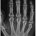
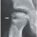
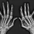
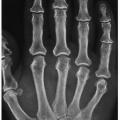
The musculoskeletal system is a common site of involvement in SLE, and joint abnormalities, exhibited by 90% of patients during the course of the disease, represent a significant part of the clinical and radiologic picture. Arthritic involvement is symmetric, and articular deformities without fixed contractures are a hallmark of this disorder. The hands are the predominant sites of involvement. Typically, the conventional radiography discloses malalignments, most commonly at the metacarpophalangeal and proximal interphalangeal joints of the fingers and the first carpometacarpal, metacarpophalangeal, and the interphalangeal joints of the thumb (Fig. 8.1). These abnormalities may not be apparent on a dorsovolar radiograph because the malalignments are flexible and are corrected by the pressure of the hand against the radiographic cassette (Fig. 8.2). These pathognomonic deformities usually occur secondary to a loss of support from the ligamentous and capsular structures about the joint and, at least in the early stage of disease, are completely reducible. Only very seldom are these abnormalities fixed
and/or accompanied by articular erosions (Fig. 8.3). Some patients present with sclerosis of the distal phalanges (acral sclerosis) (Fig. 8.4) or with resorption of the terminal tufts (acroosteolysis). Osteonecrosis, which is frequently seen, has been attributed to complications of treatment with corticosteroids (Fig. 8.5). However, current investigations suggest the vital role of the inflammatory process (vasculitis) in the development of this complication.


Figure 8.1Systemic lupus erythematosus. A: Typical appearance of the thumb in a 43-year-old woman with SLE. Note subluxations in the first carpometacarpal and metacarpophalangeal joints without articular erosions. B: In another patient, a 32-year-old woman, the oblique radiograph of her left hand shows dislocations at the first carpometacarpal joint and distal interphalangeal joint of the index finger (arrows) and subluxations in the metacarpophalangeal joints of the index and middle fingers associated with swan neck deformities (open arrows).


Figure 8.2Systemic lupus erythematosus. A: Lateral radiograph of both hands of a 42-year-old woman with documented SLE for the past 4 years demonstrates flexion deformities in the metacarpophalangeal joints. B: On the dorsovolar projection, the flexion deformities have been corrected by the pressure of the hands against the radiographic cassette.


Figure 8.3Systemic lupus erythematosus. A: A 62-year-old woman presented with a 15-year history of SLE. Dorsovolar radiograph of both hands shows severe deformities, subluxations, and articular erosions. Note the advanced osteoporosis secondary to disuse of the extremities and treatment with corticosteroids. B: In another patient, a 51-year-old woman, note flexion contractions, subluxations, and dislocations in the several joints of the right hand.


SCLERODERMA


Clinical Features

Scleroderma (progressive systemic sclerosis) is a generalized disorder of unknown cause. It is seen predominantly in young women, usually becoming apparent in their third and fourth decades. Primarily a connective tissue disorder characterized by deposition of collagen and other components of extracellular matrix in the skin and internal organs, it is distinguished by thickening and fibrosis of the skin and subcutaneous tissues, with frequent involvement of the musculoskeletal system. Scleroderma has an autoimmune basis with disease-specific ANAs, most commonly antitopoisomerase-1 and
anticentromere. Recently, researchers have identified a new genetic link to systemic form of scleroderma, a susceptibility locus involving the genome CD247 (that encodes the T-cell receptor zeta subunit modulating T-cell activation), in addition to previously known genes MHC, IRF5, and STAT4 (that encodes a regulatory protein important to immune system). However, these linkages are weak and do not provide a clear-cut genetic tool that is useful for patients. Clinically, many patients develop joint involvement, which is manifesting as arthralgia and arthropathy leading to flexion contractions of the fingers. Most patients have the so-called CREST syndrome, which refers to the coexistence of calcinosis, Raynaud phenomenon (episodes of intermittent pallor of the fingers and toes on exposure to cold, secondary to vasoconstriction of the small blood vessels), esophageal abnormalities (dilatation and hypoperistalsis), sclerodactyly, and telangiectasia; 30% to 40% of patients have a positive serologic test for rheumatoid factor and a positive antinuclear antibody (ANA) test.


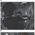
Figure 8.5SLE complicated by osteonecrosis. Oblique radiograph (A) and lateral conventional tomogram (B) of the ankle demonstrate osteonecrosis of the talus in a 26-year-old woman with lupus who was treated with massive doses of steroids. C: Coronal T2-weighted MRI in an 18-year-old woman demonstrates a focal area of osteonecrosis in the left femoral head.

Pathology shows symmetrical intimal thickening of affected arteries associated with endothelial necrosis and telangiectasia of capillaries. In the dermis, there is excessive deposition of fibrous tissue.


Imaging Features

Radiographically, scleroderma presents with characteristic abnormalities of the bone and soft tissues. The hands usually exhibit atrophy of the soft tissues at the tips of the fingers (Fig. 8.6), resorption of the distal phalanges (acroosteolysis), osteopenia, subcutaneous and periarticular calcifications (Figs. 8.7, 8.8, 8.9), and destructive changes of the small articulations, usually the interphalangeal joints (Figs. 8.9A and 8.10). Soft tissue

calcifications within the upper and lower limbs can occasionally be quite prominent (see Figs. 8.8 and 8.9B, C). Corroborative findings are seen in the gastrointestinal tract, where dilatation of the esophagus and small bowel, together with a pseudo-obstruction pattern, is characteristic (Fig. 8.11). Pseudodiverticula in the colon are also commonly seen.


Figure 8.6Scleroderma. A: A 24-year-old woman presented with atrophy of the soft tissues at the distal phalanges of the index, middle, and ring fingers (arrows). B: A radiograph of the fingers of the left hand of a 36-year-old woman shows atrophy of the soft tissue at the tip of the index and middle fingers (arrows) and early resorption of the terminal tufts (arrowheads). Compare with normal ring finger.

Only gold members can continue reading. Log In or Register to continue

Stay updated, free articles. Join our Telegram channel

Oct 1, 2018 | Posted by in GENERAL RADIOLOGY | Comments Off on Connective Tissue Arthropathies

Full access? Get Clinical Tree

Get Clinical Tree app for offline access