Chapter 36 Consequences of digital imaging in radiography
Chapter contents
36.1 Aim
The aim of this chapter is to introduce the reader to the principal consequences of digital imaging in radiography.
36.2 Image processing
36.2.1 Data clipping
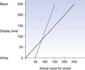
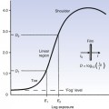
The X-ray pattern which hits the imaging plate covers a wide range of exposures. Processing all this data into an image would produce an image of very low contrast which would make image interpretation difficult. For this reason, an algorithm is applied to the image depending on the area of the body selected by the radiographer when inputting the patient data. This algorithm discards data which are not of clinical interest to the examination and so increases the contrast of the image produced. The effect of this ‘data clipping’ is shown in Figure 36.1.
36.2.2 Plate sensitivity
Once the system has clipped the data to data of clinical interest, it then has to correct the image for overall exposure. This is possible because of the linear response of the imaging plates to exposure. This is done by applying a formula which defines the average exposure within the clinically useful range. This is given a different name by different manufacturers – S number (Fuji), Exposure index (Kodak), LogM (Agfa), etc. Thus, if the plate has been overexposed, an image can be produced which has the correct density range displayed on the monitor. The principles behind this are illustrated in Figure 36.2.
Stay updated, free articles. Join our Telegram channel

Full access? Get Clinical Tree


