Chapter 1 Principles of radiography
Chapter contents
1.1 Aim
This chapter considers the basic principles of diagnostic radiography, therapeutic radiography and radiation protection. This should allow the reader to appreciate how the individual chapters within the text form part of a whole study.
1.2 Diagnostic and therapeutic radiography
1.2.1 Diagnostic radiography
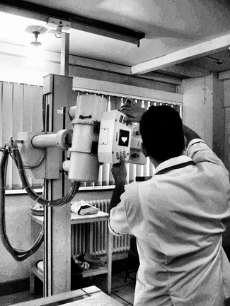
Figure 1.1 shows a radiographer preparing the equipment for a common X-ray diagnostic examination.
As light is a form of electromagnetic radiation (see Ch. 17) and it seems logical to suggest that if we can take photons of electromagnetic radiation which have higher energies than light photons, then these may have sufficient energy to penetrate body tissues and allow us to visualize internal organs. X-rays are in this part of the electromagnetic spectrum and so will penetrate body tissues and allow us to image internal organs. Unfortunately the retina of the eye cannot detect X-rays and so we cannot see an image of a structure just by directing an X-ray beam on it. This means that the X-rays, which have passed through the body, must be made to strike an image receptor that will produce a visible image, for example an imaging plate.
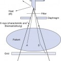
The principal interactions involved in the basic requirements for the formation of a radiographic image are shown in Figure 1.2.
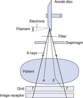
X-rays are produced in the X-ray tube by accelerating electrons and causing these to collide with the target of the X-ray tube. To understand how this works we need to know about energy (see Ch. 4) and electricity (see Chs 7 and 11) as well as the construction of the X-ray transformer and X-ray tube (see Chs 14 and 30). The X-rays so produced are over a wide band of energies. Most of the photons that have insufficient energy to be of diagnostic value are removed from the beam using filtration (see Ch. 22) and the area of the patient irradiated is restricted using a diaphragm (see Ch. 25). The beam now interacts with the patient and a number of things may happen to the X-ray photons. These may be:
• transmitted (T in Fig. 1.2): these photons pass through the patient without interacting with the patient’s tissues. They are thus unaffected by their passage through the patient
Related posts:
Stay updated, free articles. Join our Telegram channel

Full access? Get Clinical Tree


