Consolidation and Atelectasis


Consolidation and Atelectasis


W. Richard Webb


Recognizing consolidation and atelectasis is fundamental to an understanding of pulmonary radiology.


AIR-SPACE CONSOLIDATION

Air-space consolidation represents replacement of alveolar air by fluid, blood, pus, cells, or other substances. Alveolar consolidation and parenchymal consolidation are synonyms for air-space consolidation.


Radiographic Findings

Radiographic and computed tomography (CT) abnormalities indicating the presence of air-space consolidation include the following:


  • Homogeneous opacity obscuring vessels


  • Air bronchograms


  • Ill-defined or fluffy opacities


  • “Air alveolograms”


  • Patchy opacities


  • “Acinar” or air-space nodules


  • Preserved lung volume


  • Extension to the pleural surface


  • “CT angiogram” sign


Homogeneous Opacity Obscuring Vessels

With complete replacement of alveolar air, homogeneous opacification of the lung results. Vessels within the consolidated lung are invisible (Fig. 2-1A).


Air Bronchograms

In patients with consolidation, air-filled bronchi are often visible on plain radiographs or CT, appearing lucent compared with opacified lung parenchyma (see Fig. 2-1). This finding is termed an air bronchogram.

With some causes of consolidation, air bronchograms may not be visible. This usually occurs because of central bronchial obstruction (e.g., by cancer or mucus) or filling of bronchi in association with the underlying pathologic process. For example, pulmonary infarction often results in consolidation without air bronchograms because of blood filling the bronchi. In patients with bronchopneumonia, bronchi may be filled with mucus or pus.

If air bronchograms are visible within an area of consolidation, bronchial obstruction is unlikely (but not ruled out) as its cause. Although air bronchograms are considered a classic sign of air-space consolidation, they may also be seen in the presence of confluent interstitial disease.


Ill-defined or Fluffy Opacities

Consolidation often results in opacities with ill-defined margins (Figs. 2-2 and 2-3), in contrast to the relatively sharp margins of a lung mass. This results from patchy local spread of disease with variable involvement of alveoli at the edges of the pathologic process.


Air Alveolograms

If lung consolidation is not confluent, small focal lucencies representing uninvolved lung may be visible (see Fig. 2-2). These have been termed “air alveolograms,” but this is a misnomer as alveoli are too small to see radiographically. Nonetheless, these lucencies reflect incomplete lung consolidation.


Patchy Opacities

Variable consolidation in different lung regions results in patchy areas of increased opacity (see Fig. 2-3). Pulmonary vessels may be obscured or poorly defined.

Patchy consolidation visible on chest radiographs sometimes appears to be lobular or multilobular on CT (i.e., involving individual pulmonary lobules; Fig. 2-4). Some lobules appear abnormally dense while adjacent lobules appear normally aerated.


“Acinar” or Air-space Nodules

An acinus is the largest unit of lung structure in which all airways participate in gas exchange. Anatomically, it is located distal to a terminal bronchiole and is supplied by a first-order respiratory bronchiole. Acini average 7 to 8 mm in diameter.

The terms acinar nodule and air-space nodule are used to describe poorly marginated rounded opacities, usually


5 to 10 mm in diameter, that occur due to focal consolidation (Fig. 2-5). Although these nodules approximate the size of acini, they tend to be centrilobular and peribronchiolar rather than acinar. They may be seen as the only finding of consolidation or may be seen in association with larger areas of consolidation, usually at the edges of the more abnormal lung.


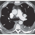
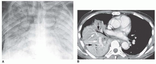
FIG. 2.1. Consolidation: homogeneous opacity obscuring vessels, air bronchograms, and the CT angiogram sign. A: Right lung consolidation due to pulmonary edema. Air bronchograms are visible bilaterally within the consolidated lung and pulmonary vessels are obscured. B: Enhanced CT in a patient with right middle and lower lobe pneumonia shows homogeneous consolidation, preserved lung volume, air bronchograms (black arrows), and opacified vessels (white arrows), appearing denser than surrounding consolidated lung (i.e., the “CT angiogram” sign).


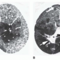
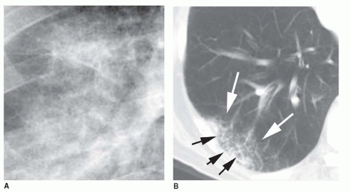
FIG. 2.2. Consolidation: ill-defined, fluffy opacities with “air alveolograms.” A: Detailed view of incomplete right lower lobe consolidation shows fluffy, ill-defined opacity containing small rounded lucencies. These lucencies have been termed air alveolograms, although they do not correspond to alveoli. B: Ill-defined fluffy consolidation (white arrows) is visible on CT in a patient with right lower lobe pneumonia. Small focal lucencies (black arrows) within the area of consolidation are “air alveolograms.”


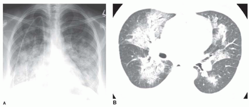
FIG. 2.3. Consolidation: patchy opacities. A: Chest radiograph in a patient with pulmonary edema due to renal failure shows patchy perihilar consolidation. B: Patchy areas of fluffy consolidation are seen on CT. The fluffy margins are due to variable involvement of alveoli at the edges of the pathologic process.


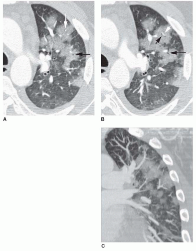
FIG. 2.4. Consolidation: patchy opacities with lobular consolidation. A, B: Contrast-enhanced HRCT in a patient with bronchopneumonia and lobular consolidation. Individual lobules are consolidated while others appear normal. Centrilobular arteries (white arrow) and bronchi (black arrows) are visible within consolidated lobules. C: Coronal reconstruction also shows the lobular distribution of the patchy lung opacities.


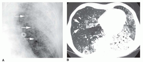
FIG. 2.5. Consolidation: acinar or air-space nodules. A: Chest radiograph shows a patchy left upper lobe pneumonia. Ill-defined nodular opacities less than 1 cm in diameter (arrows) are visible on the edge of the area of denser consolidation. These represent air-space or acinar nodules. B: CT (5-mm slice thickness) in a patient with bilateral consolidation. Air bronchograms are visible in the left lower lobe. Patchy consolidation and ground-glass opacities are present. Air-space nodules are visible in the right middle lobe (arrows) and the right lower lobe. These nodules are ill-defined, 5 to 10 mm in diameter, and centrilobular in distribution.

These nodular opacities are more easily seen on high-resolution CT (HRCT) than on chest radiographs. On HRCT, their centrilobular location is usually visible. This appearance is described further in Chapter 10.


Preserved Lung Volume

In the presence of consolidation, because alveolar air is replaced by something else (e.g., fluid), the volume of affected lung tends to be preserved (see Fig. 2-1B). Although some volume loss may be seen in patients with consolidation, it is usually of a minor degree. Alternatively, in some patients with consolidation, the lobe is expanded.


Extension to Pleural Surfaces

Pathologic processes resulting in consolidation often spread from alveolus to alveolus until reaching a fissure or pleural surface (see Fig. 2-5B). The pleural surface prevents further spread. When extension to a pleural surface occurs, the process may appear lobar, as in lobar pneumonia.


CT Angiogram Sign

A unique finding seen on CT in patients with consolidation is the “CT angiogram” sign. This sign is present if normal-appearing opacified vessels are visible within the consolidated lung following the infusion of intravenous contrast (see Figs. 2-1B and 2-6). Although opacified vessels are sometimes seen within a lung mass, they usually appear compressed or distorted.


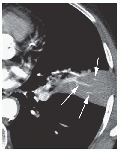
FIG. 2.6. Consolidation: the CT angiogram sign. Contrast-enhanced CT in a patient with pneumonia shows focal consolidation. Opacified arteries (arrows) appear denser than consolidated lung (i.e., the CT angiogram sign). The consolidation borders on the major fissure posteriorly and appears segmental.


Differential Diagnosis

In general, the differential diagnosis of air-space consolidation is based on a consideration of the substance that is replacing alveolar air:


  • Water (e.g., the various types of pulmonary edema)


  • Blood (e.g., pulmonary hemorrhage)


  • Pus (e.g., pneumonia)


  • Cells (e.g., bronchioloalveolar carcinoma, lymphoma, eosinophilic pneumonia, organizing pneumonia [bronchiolitis obliterans organizing pneumonia or BOOP], hypersensitivity pneumonitis)


    TABLE 2.1 Differential Diagnosis of Diffuse Consolidation


    Water (edema) (see Chapter 11)


    Hydrostatic (cardiogenic) pulmonary edema


    Heart failure


    Left atrial or pulmonary venous obstruction


    Volume overload


    Low intravascular oncotic pressure


    Hypoalbuminemia


    Liver disease


    Renal failure


    Increased permeability (noncardiogenic) pulmonary edema


    With diffuse alveolar damage (acute respiratory distress syndrome [ARDS])


    Acute interstitial pneumonia


    Aspiration of gastric acid


    Drugs


    Fat embolism


    Infection and sepsis


    Near-drowning


    Pneumonia


    Radiation


    Shock


    Toxic fumes or gases


    Trauma


    Without diffuse alveolar damage


    Any cause of ARDS, in a mild form


    Drug reactions


    Hantavirus pulmonary syndrome


    Transfusion reaction


    Mixed types of edema


    Air embolism


    High-altitude pulmonary edema


    Neurogenic pulmonary edema


    Posttransplantation edema


    Postpneumonectomy


    Reexpansion edema


    Reperfusion edema


    Tocolytic therapy


    Hydrostatic and permeability edema


    Blood (hemorrhage) (see Chapter 19)


    Aspiration of blood


    Bleeding diathesis


    Anticoagulation


    Chemotherapy


    Leukemia


    Low platelets


    Collagen-vascular disease and immune complex vasculitis


    Systemic lupus erythematosus most common


    Behçet’s syndrome


    Henoch-Schönlein purpura


    Antiphospholipid syndrome


    Goodpasture’s syndrome


    Idiopathic pulmonary hemosiderosis


    Trauma


    Vasculitis


    Wegener’s granulomatosis


    Churg-Strauss granulomatosis


    Microscopic polyangiitis


    Pus (pneumonia)


    Bacterial pneumonia


    Pneumonia in an immunosuppressed patient


    Tuberculosis


    Nontuberculous mycobacteria


    Fungal pneumonia (histoplasmosis, aspergillosis most common)


    Atypical organisms


    Virus


    Pneumocystis


    Cells


    Neoplasm


    Bronchioloalveolar carcinoma


    Lymphoma and other lymphoproliferative diseases


    Eosinophilic pneumonia or other eosinophilic diseases


    Organizing pneumonia (BOOP)


    Hypersensitivity pneumonitis


    Idiopathic interstitial pneumonias


    Nonspecific interstitial pneumonia


    Desquamative interstitial pneumonia


    Sarcoidosis


    Other substances


    Alveolar proteinosis (lipoprotein)


    Lipoid pneumonia (lipid)


  • Other substances (e.g., lipoprotein in alveolar proteinosis, lipid in lipoid pneumonia).

Patients with consolidation may be divided into two primary groups for the purpose of diagnosis: those with diffuse or bilateral consolidation and those with focal consolidation.


Diffuse Consolidation

Diffuse consolidation has a number of possible causes (Table 2-1), and the clinical history is often more important than the radiographic findings in making the diagnosis.
Nonetheless, several patterns of diffuse consolidation may suggest possible causes.


Pattern and Differential Diagnosis

Perihilarbat-wingconsolidation shows central consolidation with sparing of the lung periphery (Figs. 2-7 and 2-8). It is most typical of pulmonary edema (hydrostatic or permeability). This pattern also may be seen with pulmonary hemorrhage, pneumonias (including bacteria and atypical pneumonias such as Pneumocystis jiroveci (P. carinii) pneumonia [PCP] and viral pneumonia), and inhalational lung injury. In patients with pulmonary edema, a perihilar distribution is most often present when rapid accumulation of fluid has occurred. Relative sparing of the lung periphery has been attributed to better lymphatic clearance of edema fluid in this region, although the exact mechanism is unclear and undoubtedly varies with the disease.

Peripheral subpleural consolidation is the opposite of a bat-wing pattern (i.e., a reverse bat-wing pattern). Consolidation is seen adjacent to the chest wall, with sparing of the perihilar regions. It is most often seen in a patients with a chronic lung disease (also the reverse of what is true of a bat-wing pattern). It is classically associated with eosinophilic lung diseases, particularly eosinophilic pneumonia (Fig. 2-9A), but may also occur with organizing pneumonia (BOOP) (see Fig. 2-9B), sarcoidosis, radiation pneumonitis, lung contusion, or bronchioloalveolar carcinoma. Peripheral consolidation need not always appear peripheral on the frontal (posteroanterior [PA] or anteroposterior [AP]) radiograph; it may be peripheral in the anterior or posterior lung and overlie the parahilar regions.


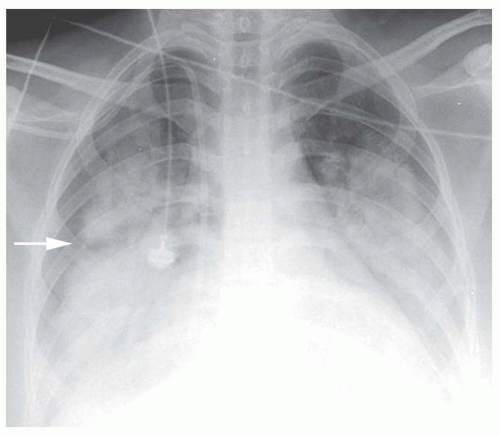
FIG. 2.7. Perihilar “bat-wing” consolidation in pulmonary edema. Chest radiograph in a patient with pulmonary edema due to renal failure (note the dialysis catheter in the right atrium) shows a distinct perihilar bat-wing pattern of consolidation. The lung periphery is spared. Note the lucency at the level of the minor fissure (arrow) because of sparing of peripheral lung adjacent to the fissure.


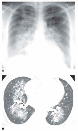
FIG. 2.8. Perihilar “bat-wing” consolidation in pulmonary edema. A: Chest radiograph shows a distinct perihilar predominance of consolidation. The heart is enlarged. B: CT shows sparing of the lung periphery.

Diffuse patchy consolidation (Fig. 2-10) may be seen with any pneumonia (bacterial, mycobacterial, fungal, viral, PCP); pulmonary edema (see Fig. 2-3A) (hydrostatic and permeability); acute respiratory distress syndrome (ARDS); pulmonary hemorrhage syndromes; aspiration; inhalational diseases; eosinophilic diseases; and diffuse bronchioloalveolar carcinoma. The patchy opacities may correspond to consolidation of lobules, subsegments, or segments.

Diffuse air-space nodules as a prominent feature of consolidation are typical of endobronchial spread of disease (Fig. 2-11). This appearance is seen in patients with endobronchial spread of infection such as tuberculosis (TB) or Mycobacterium avium complex (MAC), bacterial bronchopneumonia,
viral pneumonia (cytomegalovirus [CMV], measles), endobronchial spread of bronchioloalveolar carcinoma, pulmonary hemorrhage, or sometimes aspiration.


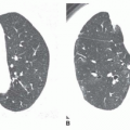
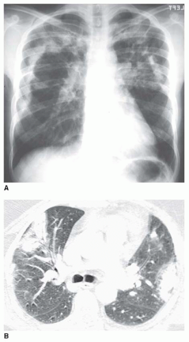
FIG. 2.9. Peripheral subpleural (reverse bat-wing) consolidation. A: Chest radiograph in a patient with chronic eosinophilic pneumonia shows areas of consolidation in the subpleural lung. The perihilar regions are spared. B: CT in a patient with BOOP shows patchy areas of consolidation in the subpleural lung.

Diffuse homogeneous consolidation is most typical in patients with pulmonary edema, ARDS, pulmonary hemorrhage, pneumonias (including viral and PCP), alveolar proteinosis, and extensive atelectasis.


Focal Consolidation

The most likely causes of focal consolidation include pneumonia, atelectasis with or without bronchial obstruction, and neoplasm, while pulmonary edema and hemorrhage are much less likely than in patients with diffuse abnormalities.


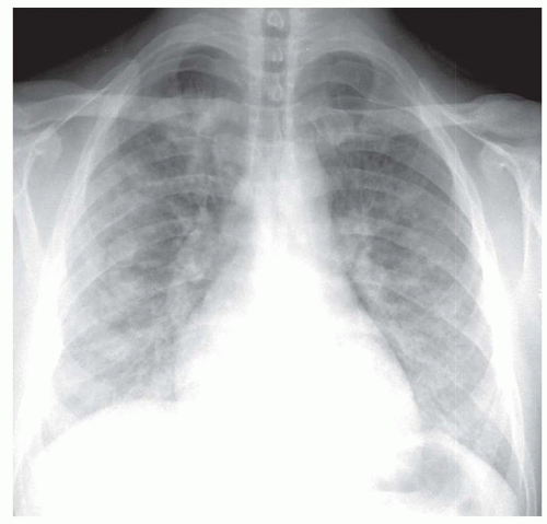
FIG. 2.10. Diffuse patchy consolidation in a patient with viral pneumonia.


Pattern and Differential Diagnosis

Focal consolidation may represent pneumonia; postobstructive pneumonia; aspiration; bronchioloalveolar carcinoma; lymphoma or other lymphoproliferative disease; infarction; hemorrhage due to trauma, pulmonary embolism, or diseases such as Wegener’s granulomatosis; pulmonary infarction; radiation pneumonitis; organizing pneumonia (BOOP);
eosinophilic pneumonia; atelectasis; or rarely focal edema. The appearance of focal consolidation may also result from confluent interstitial disease, as in patients with sarcoidosis. The appearance or pattern of focal or multifocal consolidation may be helpful in differential diagnosis.


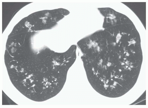
FIG. 2.11. Diffuse air-space nodules in bronchopneumonia. Multiple small nodular opacities are typical of spread of infection through the airways. This represented a bacterial bronchopneumonia, but other organisms such as TB, MAC, fungus, or viruses may be involved.


TABLE 2.2 Differential Diagnosis of Focal Consolidation


Water (edema) (uncommon)


Papillary muscle rupture with mitral prolapse (right upper lobe)


Edema in a patient with


Pulmonary artery obstruction (e.g., pulmonary embolism)


Hypoplastic pulmonary artery


Swyer-James syndrome


Decubitus position


Reexpansion edema


Pulmonary vein occlusion


Systemic to pulmonary artery shunt (congenital or acquired)


Bland aspiration


Atelectasis with drowned lung


Blood (hemorrhage)


Contusion


Infarction


Aspiration of blood


Vasculitis


Pus (pneumonia)


Bacterial


Tuberculosis or nontuberculous mycobacterial


Fungal


Virus (uncommon)


Pneumocystis (uncommon)


Aspiration pneumonia


Atelectasis with postobstructive pneumonia


Cells


Neoplasm


Bronchioloalveolar carcinoma


Lymphoma and other lymphoproliferative diseases


Eosinophilic pneumonia or other eosinophilic diseases


Organizing pneumonia (BOOP)


Sarcoidosis


Other substances


Lipoid pneumonia (lipid)


Lobar consolidation is most typical of pneumonia (including S. pneumoniae, Klebsiella (Fig. 2-12A), Legionella, and TB) and abnormalities associated with bronchial obstruction.

Lobar consolidation with pneumonia often occurs because of interalveolar spread of disease via the pores of Kohn (small holes in the alveolar walls). This type of spread continues until a fissure or pleural surface is reached. Organisms associated with spread of pneumonia via the pores of Kohn are characterized by thin secretions (thus passing easily through the pores). The presence of an incomplete fissure may lead to a lobar pneumonia becoming bilobar (or trilobar) (see Fig. 2-1B).


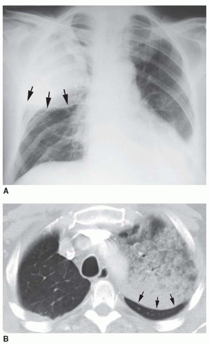
FIG. 2.12. Lobar consolidation with expansion. A: A patient with right upper lobe consolidation due to Klebsiella pneumonia shows downward bowing of the minor fissure (arrows) because of lobar expansion. B: Bronchioloalveolar carcinoma involving the left upper lobe with posterior bulging (arrows) of the left major fissure.

Lobar consolidation resulting from interalveolar spread of disease can also be seen with lymphoma and bronchioloalveolar carcinoma (see Fig. 2-12B). The term lepidic growth is used to describe the local interalveolar spread of tumors such as bronchioloalveolar carcinoma, using alveolar walls as a scaffold.

Bronchial obstruction with postobstructive pneumonia or atelectasis also commonly results in lobar consolidation. The differential diagnosis is that of bronchial obstruction and includes neoplasm, lymph node enlargement, inflammatory lesions, stricture, and foreign body.

Lobar consolidation is uncommonly the result of vascular abnormalities. Right upper lobe consolidation representing pulmonary edema may occur in patients with acute
myocardial infarction resulting in papillary muscle rupture and mitral valve prolapse; it occurs because a jet of regurgitant blood is directed into the right superior pulmonary vein. Focal pulmonary hemorrhage may lead to a lobar consolidation. Lobar consolidation is uncommon with pulmonary embolism.

Lobar expansion in association with lobar consolidation suggests infection, particularly by Klebsiella (see Fig. 2-12A) or Pneumococcus, TB, bronchial obstruction with postobstructive pneumonia, or consolidation associated with neoplasm (see Fig. 2-12B).


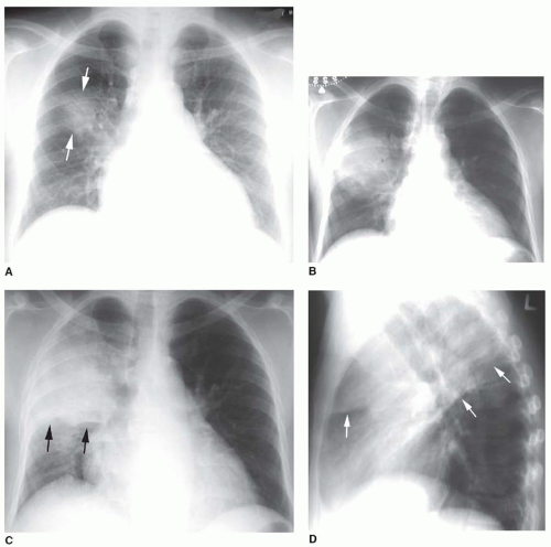
FIG. 2.13. Spherical consolidation due to pneumonia. A: On the initial radiograph, a patient with Legionella pneumonia shows a poorly defined area of consolidation (arrows) in the right upper lobe. This may be termed “round pneumonia.” B: Over the next several days, the spherical consolidation increases in size because of local interalveolar spread. This appearance may be seen in the early stages of lobar pneumonias. C: Further progression results in consolidation of the right upper lobe, marginated by the minor fissure (arrows). D: A lateral view at the same time as C shows upper lobe consolidation marginated by the major and minor fissures (arrows). Partial right middle lobe consolidation is also present.

Round or spherical consolidation is most typical of bronchioloalveolar carcinoma, lymphoma or lymphoproliferative disease, or pneumonia (i.e., round pneumonia). A round or spherical pneumonia is typical of organisms that spread via the pores of Kohn and progress to being lobar, such as S. pneumoniae, Klebsiella, Legionella, or TB (Fig. 2-13). Such diseases begin in at a single site and result in an enlarging ill-defined
sphere of consolidation as more and more alveoli become involved. As the growing sphere reaches a pleural surface or fissure and cannot spread further, it becomes lobar.

Segmental (or subsegmental) consolidation may be diagnosed if a wedge-shaped opacity of more than a few centimeters in size is visible with the apex of the wedge pointing toward the hilum (see Figs. 2-6 and 2-14). This finding suggests an abnormality related to a segmental (or subsegmental) bronchus or artery, such as bronchial obstruction due to mucus or tumor, bronchopneumonia, focal aspiration, or pulmonary embolism with infarction.

Focal patchy consolidation is typical of pneumonias, endobronchial spread of TB, or endobronchial spread of tumor such as bronchioloalveolar carcinoma (see Fig. 2-2). CT may show a pattern of lobular consolidation. Centrilobular nodules are seen in some cases (see Fig. 2-4).

Patchy consolidation is typical of bronchopneumonia. Pneumonias associated with this pattern (e.g., Staphylococcus, Haemophilus, Pseudomonas) are characterized by thick and tenacious secretions and spread via airways rather than the pores of Kohn. Infected secretions are typically present within the bronchi. Bronchopneumonia is also known as lobular pneumonia because of its tendency to involve individual lobules. Mycoplasma pneumonia often results in this pattern.


Time Course in Diagnosis

Rapidly appearing consolidation (a few hours) suggests atelectasis with drowned lung, aspiration, pulmonary edema, pulmonary hemorrhage, infarction, or rapidly progressing pneumonia, particularly in an immunocompromised host. Of these, only pulmonary edema and drowned lung may clear quickly. Occasionally, a lymphoproliferative neoplasm progresses within hours.


FIG. 2.14. Segmental consolidation. A patient with pneumonia shows consolidation of the lateral segment of the right middle lobe. The segmental bronchus is seen within the consolidated lung as an air bronchogram. The medial segment, adjacent to the right heart border, is normally aerated. The consolidated segment borders posteriorly on the major fissure.

Longstanding (chronic) consolidation (4 to 6 weeks) with little change suggests eosinophilic pneumonia, BOOP, bronchioloalveolar carcinoma, lymphoma, lipoid pneumonia, or some indolent pneumonias such as fungal infections. Recurrent processes (e.g., recurrent pulmonary edema, pulmonary hemorrhage, or aspiration) may appear to be chronic if radiographs are obtained only during the acute episodes.


SILHOUETTE SIGN

The borders of soft tissue structures such as the mediastinum, hila, and hemidiaphragms are visible on chest radiographs because they are outlined by adjacent air-containing lung. When consolidated lung (or a soft tissue mass) contacts one of these structures, its border becomes invisible or is poorly marginated. This is termed the “silhouette” sign. The silhouette sign is used to diagnose the presence of a lung abnormality (i.e., consolidation, atelectasis, mass) and localize it to a specific lobe or lung region (Figs. 2-15,2-16,2-17,2-18,2-19 and 2-20).

On the frontal (PA or AP) radiograph, obscuration (in radiologic parlance, “silhouetting”) of specific contours may be related to abnormalities in specific lobes. Specific contours and their corresponding lobes are illustrated in Figure 2-15 and listed below:

Right superior mediastinum (i.e., superior vena cava [SVC]) = right upper lobe (see Fig. 2-16)

Right heart border = right middle lobe (common; see Fig. 2-17) or medial right lower lobe (less common). This appearance may be mimicked by pectus excavatum.

Right hemidiaphragm = right lower lobe (see Fig. 2-18)

Left superior mediastinum (e.g., aortic arch) = left upper lobe (see Fig. 2-19B)

Left heart border = lingular segments of left upper lobe (see Fig. 2-19)

Left hemidiaphragm or descending aorta = left lower lobe (see Fig. 2-20)

Caveat: The diaphragmatic contour seen on the frontal (PA or AP) radiograph represents the dome, or the highest point, of the diaphragm. The diaphragmatic dome is relatively anterior, and lower lobe consolidation may be posterior to it (see Fig. 2-21); in this case, the hemidiaphragm may remain visible.

The silhouette sign is also used on the lateral projection:

Posterior margin of the heart or posterior left hemidiaphragm = left lower lobe (see Fig. 2-20); hiatal hernia may mimic this.

Anterior right hemidiaphragm = right middle lobe

Posterior right hemidiaphragm = right lower lobe (see Figs. 2-18 and 2-21)


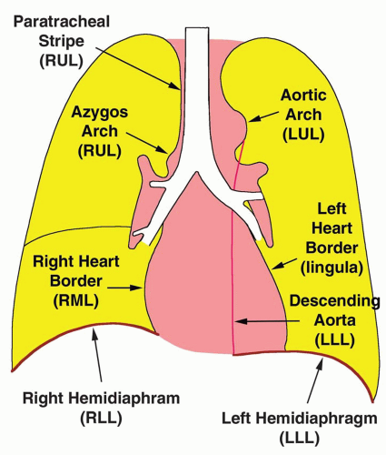
FIG. 2.15. Anatomic relationships used with the silhouette sign. Obscuration of the borders shown in this diagram are associated with consolidation of the listed lobes. RUL, right upper lobe; RML, right middle lobe; RLL, right lower lobe; LUL, left upper lobe; LLL, left lower lobe.


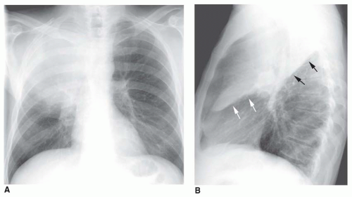
FIG. 2.16. The silhouette sign in right upper lobe pneumonia. A: Consolidation of the right upper lobe obscures (i.e., silhouettes) the border of the right superior mediastinum and superior vena cava. The upper part of the right hilum is also invisible. B: On the lateral view, the consolidated upper lobe is outlined superiorly by the upper aspect of the major fissure (black arrows). Inferiorly, it is outlined by the minor fissure (white arrows). (Figure continues.)


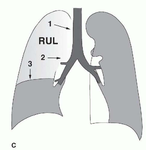
FIG. 2.16. (Continued.) C: Typical findings of right upper lobe consolidation: (1) obscuration of the right superior mediastinum, (2) obscuration of the superior right hilum, and (3) opacity marginated inferiorly by the minor fissure.

Only gold members can continue reading. Log In or Register to continue

Stay updated, free articles. Join our Telegram channel

Oct 10, 2016 | Posted by in CARDIOVASCULAR IMAGING | Comments Off on Consolidation and Atelectasis

Full access? Get Clinical Tree

Get Clinical Tree app for offline access