Lymphoma and Lymphoproliferative Disease


Lymphoma and Lymphoproliferative Disease


W. Richard Webb


Lymphoma accounts for about 4% of newly diagnosed malignancies and commonly involves the mediastinum, hila, and lung parenchyma.

Lymphomas are primary neoplasms of the lymphoreticular system and are classified in two main types: Hodgkin’s disease (HD) and non-Hodgkin’s lymphoma (NHL). Although HD is the less common of the two types, representing about 25% to 30% of cases, it is more common as a cause of mediastinal disease.


HODGKIN’S DISEASE

HD occurs at all ages, but its peak incidence is in the third and eighth decades; it accounts for about 0.5% to 1% of all newly diagnosed malignancies (Table 5-1). It is more prevalent in males, with a male-to-female ratio ranging from 1.4 to 1.9. Intrathoracic HD usually is associated with disease elsewhere; cervical lymph nodes are commonly involved. Constitutional symptoms may be present.

HD is characterized histologically by the presence of Reed-Sternberg cells. Four histologic types of HD are recognized in the Rye classification: nodular sclerosis (accounting for 50% to 80% of adult HD cases); lymphocyte predominance; mixed cellularity; and lymphocyte depletion.

HD has a predilection for thoracic involvement. Up to 85% of patients with HD have thoracic involvement at the time of diagnosis; nearly all of them have mediastinal lymph node enlargement.


Lymph Node Involvement

HD most often involves superior mediastinal (i.e., prevascular, paratracheal, and aortopulmonary) lymph nodes. These node groups are abnormal in as many as 85% of patients with HD and 98% of those with thoracic involvement (Figs. 5-1 and 5-2); if these nodes appear normal on computed tomography (CT), intrathoracic adenopathy is unlikely to represent HD.

Other sites of involvement in patients with thoracic disease are as follows: the hilar nodes, in about 35% of patients; the subcarinal nodes, in about 25%; the paracardiac (cardiophrenic angle) lymph nodes, in 10%; the internal mammary nodes, in 5%; and the posterior mediastinal (i.e., paravertebral, paraaortic, and retrocrural) nodes, in 5% (see Figs. 5-2 and 5-3).

Multiple node groups are involved in 85% of those HD patients who have thoracic node involvement. Enlargement of a single node group can be seen in some patients with HD, but it is uncommon, occurring in only 15% of cases with node involvement. Anterior (prevascular) lymph nodes are most often involved as a single group (Fig. 5-4), and this appearance usually indicates the presence of nodular sclerosing HD.

On plain radiographs, anterior mediastinal lymph node enlargement may result in a unilateral or bilateral mediastinal abnormality (see Figs. 5-1A and 5-3A). Enlargement of paratracheal or aortopulmonary window nodes often results in a unilateral or asymmetrical abnormality. Because

multiple lymph nodes are involved, mediastinal masses in HD often appear elongated or lobulated in contour. Roughly spherical masses also can be seen (Fig. 5-5). Poor definition of the mass can indicate invasion or extension into adjacent lung.


TABLE 5.1 Hodgkin’s Disease


Peak incidence in the third and eighth decades


Characterized by Reed-Sternberg cells


Nodular sclerosis cell type accounts for 50%-80% of adult cases


Staged using Ann Arbor system


Thoracic involvement in 85% of cases at diagnosis


Lymph node involvement in nearly all with thoracic involvement


Superior mediastinal node enlargement in 98%


Multiple node groups involved in 85%


Nodes show low attenuation in 10%-20%


Residual mediastinal masses common after treatment


Calcification of lymph nodes common after treatment


Lung disease


10% have lung involvement at diagnosis


Nearly always associated with enlarged nodes


Direct infiltration, lung nodules, or consolidation


Air bronchograms and cavitation may be seen


Lung recurrence may occur without enlarged nodes


Pleural effusion in 15%, usually due to lymphatic obstruction


FIG. 5.1. HD involving superior mediastinal lymph nodes. A: PA chest radiograph shows bilateral lobulated superior mediastinal masses (arrows). B: The lateral view shows an anterior mediastinal mass (arrows). C: Contrast-enhanced CT scan shows prevascular anterior mediastinal lymph node enlargement (arrows) and pretracheal lymph node enlargement (*). Lymph node enlargement in these regions is typical of HD.


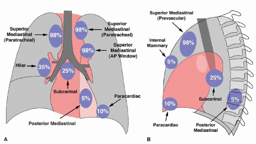
FIG. 5.2. Involvement of mediastinal lymph node groups in HD, illustrated as a percentage of patients with thoracic disease. Lymph node groups as shown on the PA (A) and lateral (B) radiographs.


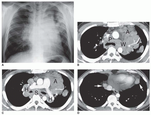
FIG. 5.3. HD with involvement of multiple lymph node groups. A: PA chest radiograph shows superior mediastinal widening, hilar enlargement, and nodules in the left upper lobe. B, C: Large lymph nodes are visible in the prevascular anterior mediastinum (A), pretracheal space (P), aortopulmonary window (W), subcarinal space (S), and both hila (H). Some discrete lymph nodes are visible, but other enlarged node masses appear matted together, with fat planes between them being invisible. D: Paracardiac lymph node enlargement (arrows) is visible at a lower level on CT.


FIG. 5.4. Anterior mediastinal lymph node enlargement in nodular sclerosing HD. CT shows lymphadenopathy localized to the anterior mediastinum, which is typical of nodular sclerosing HD.

CT is advantageous in showing abnormalities of mediastinal lymph nodes in patients with HD. Although it is uncommon for CT to show evidence of mediastinal adenopathy if the chest radiograph is normal, in cases in which the radiograph shows lymph node enlargement, CT detects additional sites of adenopathy in many patients. Findings shown only on CT may change the treatment plan in as many as 10% of patients. CT is most helpful in diagnosing subcarinal, internal mammary, and aortopulmonary window node enlargement that is not visible on radiographs.

On CT, abnormal lymph nodes may appear welldefined and discrete (Fig. 5-6), may appear matted (with

intervening fat planes poorly seen; see Fig. 5-3B and C), or may be associated with diffuse mediastinal infiltration (with individual lymph nodes being invisible; see Fig. 5-5C and D). Most often, enlarged lymph nodes are of homogeneous soft tissue attenuation, but in 10% to 20% of cases, lymph node masses show areas of low attenuation or necrosis following contrast enhancement (Fig. 5-7). Inhomogeneity without obvious necrosis also may be seen (see Fig. 5-5D). Invasion of mediastinal structures such as the superior vena cava, esophagus, or airways may occur.


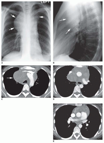
FIG. 5.5. Large mediastinal mass in HD. A: The PA radiograph shows a large spherical mass projecting to the right, with a smaller left-sided component (arrows). B: The lateral view shows the mass to be anterior (arrows). C: At the level of the aortic arch, CT shows a large, rounded mass that largely involves the pretracheal mediastinum (arrow). The opacified brachiocephalic veins are displaced anteriorly. Discrete nodal masses are not visible; the mediastinum appears infiltrated by tumor, and no mediastinal fat is visible. D: At the level of the left pulmonary artery, CT reveals that the mass occupies the prevascular anterior mediastinum and the precarinal space. The superior vena cava is displaced anteriorly and is compressed. At this level the mass appears somewhat inhomogenous in attenuation. Discrete, enlarged lymph nodes are not seen. E: At the level of the right pulmonary artery, anterior mediastinal mass appears to represent thymic involvement.


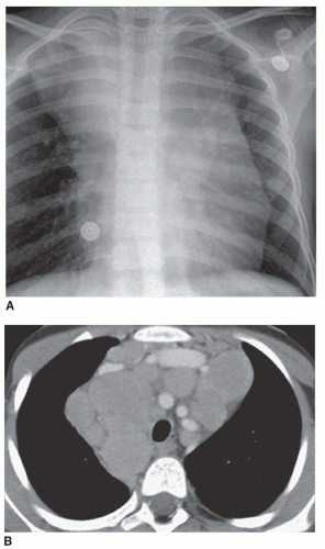
FIG. 5.6. HD in a 9-year-old child. A: Chest radiograph shows bilateral superior mediastinal masses. B: On CT, multiple discrete, enlarged lymph nodes are visible in the middle and anterior mediastinum.

Rarely, untreated patients show fine, stippled lymph node calcification. Lymph node calcification is much more common following treatment, with a stippled, confluent, or, less often, “egg-shell” appearance (Fig. 5-8). Calcification usually occurs after radiation; calcification after chemotherapy is less common.


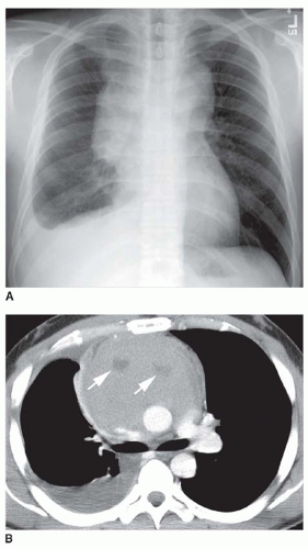
FIG. 5.7. Lymphoma with necrosis. A: Chest radiograph shows a large bilateral mediastinal mass and right pleural effusion, a portion of which is subpulmonic. B: Contrast-enhanced CT scan shows an anterior mediastinal mass containing an area of low attenuation (arrows). This finding is seen in 10% to 20% of patients with HD.

HD also has a predilection for involvement of the thymus in association with mediastinal lymph node enlargement. Thymic enlargement is seen in 30% to 40% of cases, but may be difficult to distinguish from an anterior mediastinal lymph node mass unless the normal thymic shape is preserved (see Figs. 5-5E and 5-9). In the presence of thymic involvement, a visible mediastinal mass can project to both sides of the mediastinum.


FIG. 5.8. Lymph node calcification following radiation treatment for HD. A: Coned-down lateral radiograph shows stippled calcification of enlarged anterior mediastinal lymph nodes (arrows), typical of radiated HD. Enlarged residual lymph nodes commonly are seen after treatment of HD. B, C: Focal calcifications of enlarged anterior mediastinal lymph nodes are shown on CT. Residual mediastinal lymph node masses following treatment of lymphoma are common, and most typical of nodular sclerosing HD.


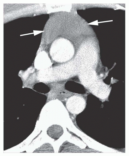
FIG. 5.9. Thymic involvement in HD. The thymus is enlarged but maintains a normal shape (arrows).

HD is believed to be unifocal in origin, spreading to involve contiguous lymph nodes. It is unusual for HD to skip lymph node groups, and if nodes contiguous with the mediastinum, such as the lower neck or upper abdomen, are not involved by HD, it usually is not necessary to scan more distant regions, such as the pelvis.

However, in patients with mediastinal HD, scanning always should be extended to include the upper abdomen. Intra-abdominal paraaortic adenopathy can be found in 25% of patients with HD, and the spleen and liver are involved in 35% and 10% of patients, respectively.

The magnetic resonance imaging (MRI) appearance of lymph node masses in HD varies with the histology. In nodular sclerosing HD, large amounts of fibrous tissue typically are interlaced with malignant cells. Patients typically show a heterogeneous pattern with mixed high and low signal intensities on T2-weighted images. Low signal intensity areas on T2-weighted images are related to regions of fibrosis in the tumor, and high-intensity regions represent tumor tissue or cystic regions. HD also may demonstrate homogeneous high signal intensity similar to that of fat on T2-weighted images.


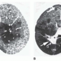
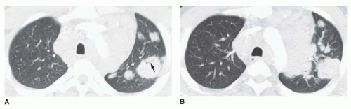
FIG. 5.10. Lung involvement in HD in the same patient shown in Figure 5-3. A, B: Large lobulated nodules are visible in the left lung. Some are associated with small bronchi or contain air bronchograms (arrow). Extensive mediastinal and hilar lymph node enlargement is present.


Lung Involvement

Lung involvement by HD is seen in 10% of patients at the time of presentation. It is almost always associated with mediastinal (and usually ipsilateral hilar) adenopathy (Fig. 5-10). A variety of manifestations of lung involvement may be seen, but the most common are (a) direct invasion of lung contiguous with abnormal nodes and (b) isolated single or multiple lung nodules, masses, or areas of consolidation. Direct invasion and the presence of nodules or masses occur with about equal frequency.

Direct extension from hilar or mediastinal nodes results in coarse linear or streaky opacities radiating outward into the lung, corresponding on CT to thickening of the peribronchovascular interstitium. Interlobular septal thickening may be associated. In some patients, the appearance may mimic that of lymphangitic spread of carcinoma.

Discrete, single or multiple, well-defined or ill-defined, large or small lung nodules or mass-like lesions, or localized areas of air-space consolidation associated with air bronchograms may be seen (Figs. 5-10 and 5-11). These can cavitate, with thick or thin walls. Peripheral, subpleural masses are relatively common (Fig. 5-12).


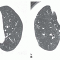
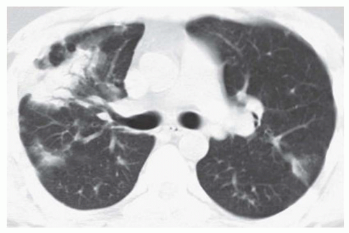
FIG. 5.11. Lung involvement in recurrent HD. Poorly defined nodules and areas of consolidation are visible. The large area of consolidation on the right contains a number of air bronchograms. There is no obvious lymph node enlargement.

HD occasionally involves bronchi with endobronchial masses or bronchial compression associated with atelectasis (Fig. 5-13).

In previously untreated patients, lung disease is uncommon in the absence of radiographically demonstrable lymph node enlargement; however, lung recurrence can be seen without node enlargement in patients with prior mediastinal radiation (see Fig. 5-11).


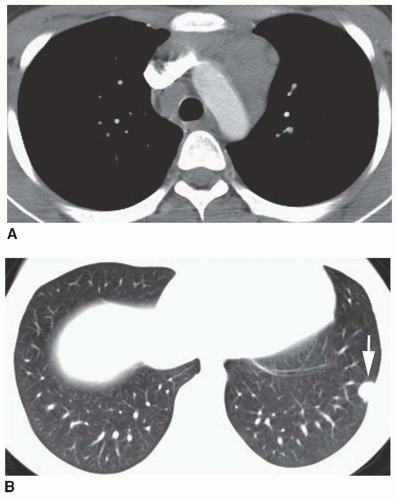
FIG. 5.12. Peripheral lung nodule in HD. A: CT shows pretracheal and prevascular lymph node enlargement. B: A small, well-defined subpleural nodule is visible in the left lung. This was found at biopsy to represent HD.


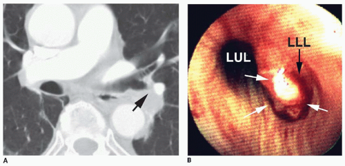
FIG. 5.13. Endobronchial HD. A: The left lower lobe bronchus is narrowed (arrow) by a polypoid endobronchial mass. B: Endoscopic photograph showing the left upper lobe bronchus (LUL) and a polypoid mass (white arrows) filling the left lower lobe bronchus (LLL).


Pleural and Pericardial Effusion

Pleural effusion is present in about 15% of patients at diagnosis and usually reflects lymphatic or venous obstruction rather than pleural involvement by tumor (see Fig. 5-7). Effusions tend to resolve following local mediastinal or hilar radiation. However, about 20% to 25% of patients with HD who have effusion have CT findings of pleural or extrapleural tumor or lymph node enlargement. Pericardial effusion, present in 5% of patients, usually indicates direct involvement of the pericardium.


TABLE 5.2 Ann Arbor Staging Classification for Lymphoma


Stagea


Definition


I


Involvement of a single lymph node region (I) or a single extralymphatic organ or site (IE)


II


Involvement of two or more lymph node regions on the same side of the diaphragm (II) or localized involvement of an extralymphatic organ or site and of one or more lymph node regions on the same side of the diaphragm (IIE)


III


Involvement of lymph node regions on both sides of the diaphragm (III), which may also be accompanied by involvement of the spleen (IIIS) or by localized involvement of an extralymphatic organ or site (IIIE) or both (IIISE)


IV


Diffuse or disseminated involvement of one or more extralymphatic organs or tissues, with or without associated lymph node involvement


a The absence or presence of fever, night sweats, and/or unexplained loss of 10% or more of body weight in 6 months is denoted by the suffix A or B, respectively.


Chest Wall Involvement

Invasion of the chest wall contiguous with mediastinal or lung masses occurs in about 5% of cases. Tumor may involve ribs, sternum, or vertebral bodies and typically results in lytic bone destruction. Involvement of the skeleton because of dissemination often results in mixed lytic and blastic lesions (e.g., “ivory vertebra”).


Staging

The Ann Arbor Staging Classification is used to describe the anatomic extent of the disease at the time of diagnosis and correlates well with prognosis (Table 5-2). Radiation is used for treatment of stages I and II. A combination of radiation and chemotherapy or chemotherapy alone is used in stages III and IV. There is a 75% to 80% cure rate for adult HD; in children, the cure rate is about 95%.


LYMPHOMA TREATMENT RESPONSE AND RELAPSE

Imaging studies commonly are obtained to judge the completeness of tumor response to treatment and to diagnose relapse.

Reduction of tumor bulk is always seen in patients with adequately treated tumor. Patients who show complete resolution of lymph node masses on CT usually are considered to have had a satisfactory response.


Residual mediastinal mass or lymph node enlargement often is seen in cured patients following treatment (see Figs. 5-8 and 5-14). This is particularly common in patients with treated nodular sclerosing HD; residual masses represent fibrous tissue components of the tumor itself, which change little with treatment, or post-treatment fibrosis. Residual mediastinal masses can be seen in as many as 88% of patients with HD and 40% of patients with NHL. In most patients, masses remain unchanged on follow-up. In some patients, residual masses continue to decrease in size or resolve over a period ranging from 3 to 11 months.


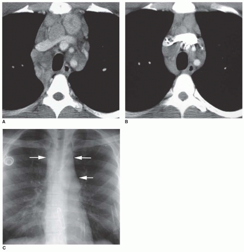
FIG. 5.14. Residual mediastinal mass in HD. A: CT scan before treatment shows multiple enlarged mediastinal lymph nodes. B: CT performed 2 years after treatment shows a decrease in lymph node size, but lymph nodes remain enlarged. Persistent enlarged lymph nodes are common after treatment of HD. C: Chest radiograph taken at the same time as the CT scan shown in (B) shows widening of the superior mediastinum and prominence of the aortopulmonary window (arrows).

Most patients with thymic enlargement resulting from HD show a return to normal thymic size following treatment, although residual thymic enlargement may be seen in about 30% of cases.

Recurrent HD does not commonly involve previously irradiated (i.e., in field) intrathoracic lymph nodes. However, so-called “in-field recurrence” is seen in a small percentage of cases. Large masses and masses in the anterior mediastinum generally are considered to carry an increased risk of recurrence.

Common sites of recurrent HD include the upper mediastinum and lung, both seen in about half of cases with
recurrence. Lymph node recurrence also may involve paracardiac lymph nodes, which usually are excluded from the treatment field because of their close relation to the heart (to avoid radiation pericarditis). Lung recurrence usually is associated with central or peripheral lung nodules, masses, or areas of focal consolidation, any of which may show cavitation. Lung recurrence is uncommonly associated with visible lymph node enlargement on chest radiographs, although it is visible on CT in about half of cases. Pleural effusions or masses and chest wall involvement also are common.


TABLE 5.3 Non-Hodgkin’s Lymphoma


Diverse group of neoplasms


Specific tumor types classified by WHO


More common than HD


Mean age 55 years; more common than HD in children


Associated with immunodeficiency, HIV, immunosuppression


Prognosis related to cell type


Staging less important than with HD


Thoracic involvement in 40%-50%


Lymphadenopathy in 75% with thoracic disease


Superior mediastinal lymph nodes involved in nearly all


Involvement of a single node group common (40%)


Posterior mediastinal lymph nodes relatively common


Lung involvement (30%) more common than with HD


A homogenous hypointense pattern on both T1- and T2-weighted MRI images is characteristic of residual fibrotic masses in cured patients. Approximately 80% of cases show this pattern within 6 to 8 weeks of treatment. A heterogeneous appearance on T2-weighted images is seen after treatment in about 20% of cases, with regions of high signal intensity representing areas of necrosis or inflammation, or, in some patients, residual tumor. Thus, a high relative intensity with T2 weighting indicates the need for biopsy, follow-up, or further imaging. Gallium-67 or positron emission tomographic (PET) imaging can help in distinguishing active tumor from residual benign masses.


TABLE 5.4 Comparison of HD and NHL

Only gold members can continue reading. Log In or Register to continue

Stay updated, free articles. Join our Telegram channel

Oct 10, 2016 | Posted by in CARDIOVASCULAR IMAGING | Comments Off on Lymphoma and Lymphoproliferative Disease

Full access? Get Clinical Tree

Get Clinical Tree app for offline access