The Trachea


The Trachea


W. Richard Webb


THE NORMAL TRACHEA

The trachea extends from the inferior aspect of the cricoid cartilage (at the level of the sixth cervical vertebra) to the carina (at the level of the fifth thoracic vertebra). It measures from 10 to 12 cm in length. The trachea is divided into extrathoracic and intrathoracic portions at the level it passes posterior to the manubrium; the extrathoracic trachea is 2 to 4 cm in length, while the intrathoracic trachea measures 6 to 9 cm in length. From 16 to 22 horseshoe-shaped bands of hyaline cartilage support the anterior and lateral tracheal walls. The incomplete posterior portion of these rings is bridged by a thin band of smooth muscle and fibrous tissue, the posterior tracheal membrane (Fig. 22-1).

The plain film appearance of the trachea is described in Chapter 7. On CT, the trachea usually is round or oval in shape, but it can appear horseshoe-shaped, triangular, or like an inverted pear in some normal patients. The tracheal wall is delineated by air in its lumen internally and by mediastinal fat externally, and is usually visible as a 1- to 2-mm soft tissue stripe. The posterior tracheal membrane appears thinner than the anterior and lateral tracheal walls, and is variable in shape due to its lack of cartilage; it can appear convex, concave, or flat. The tracheal cartilages may appear calcified or slightly denser than adjacent soft tissue. Calcification of cartilage is most common in older patients, and is particularly common in women (Fig. 22-2). In patients with calcified cartilage, little soft tissue is seen in the tracheal wall internal to the cartilage.

Tracheal diameter varies widely in normal subjects. In normal men, tracheal diameter averages 19.5 mm, with a range of 13 to 25 mm (mean ± 3 SD) in the coronal plane and 13 to 27 mm in the sagittal plane. In women, tracheal diameter is slightly smaller, averaging 17.5 mm and ranging from 10 to 21 mm in the coronal plane and 10 to 23 mm in the sagittal plane.

On CT performed during or after forced expiration, the posterior tracheal membrane bulges anteriorly, narrowing and, in some cases, nearly obliterating the tracheal lumen (Fig. 22-3). The mean anterior-posterior diameter of the trachea decreases by 30% to 40% during forced expiration due to anterior bulging of the posterior membrane; the transverse diameter decreases by 10% to 20%. The cross-sectional area of the trachea may diminish by more than 50% in normal subjects with forced expiration.


Tracheal Bronchus

A tracheal bronchus represents the origin of all or part (usually the apical segment) of the right upper lobe bronchus from the trachea; its incidence is about 0.1% (see Figs. 1-1 and 1-2 in Chapter 1). Left tracheal bronchus also occurs but is much less common.


ABNORMALITIES ASSOCIATED WITH TRACHEAL NARROWING

Focal tracheal narrowing may be seen with tracheal tumors, tuberculosis (TB), tracheal stenosis or stricture, tracheomalacia, or occasionally with saber sheath trachea, Wegener’s granulomatosis, or amyloidosis. Diffuse or generalized tracheal narrowing may be seen with tracheomalacia, saber sheath trachea, Wegener’s granulomatosis, amyloidosis, tracheobronchopathia osteochondroplastica, and relapsing polychondritis.


Tracheal Tumors

Tumors of the trachea are rare (Table 22-1). Symptoms often are absent or nonspecific (e.g., cough, dyspnea), and early diagnosis is difficult. Tracheal tumors tend to be inconspicuous on chest radiographs, and may become quite large before they are detected. CT is highly sensitive in detecting tracheal tumors and their extent of spread. Together, squamous cell carcinoma and adenoid cystic carcinoma account for more than 85% of tracheal tumors. Tumors of many other cell types, both epithelial and mesenchymal, may occur in the trachea but are much less common.

Squamous cell carcinoma is associated with smoking and is multifocal in 10% of cases, often involving the distal trachea; a main bronchus also may be involved (see Figs. 3-19 and 3-20 in Chapter 3). Adenoid cystic carcinoma originates from tracheal mucous glands and is most common in the upper trachea (Figs. 22-4 and 22-5). It is slightly less common than squamous cell carcinoma. Adenoid cystic carcinoma often arises from the posterolateral tracheal wall (Figs. 22-5 and 22-6B).

On CT, a primary malignant tracheal tumor may appear as a polypoid lesion, a focal sessile lesion, eccentric narrowing of the tracheal lumen, or circumferential wall thickening
(see Fig. 22-5). Attachment to the tracheal wall may be either broad based (Fig. 22-5) or narrow and pedunculated. CT may underestimate the longitudinal extent of the tumor; submucosal spread may be difficult to see on CT. However, CT is superior to bronchoscopy in evaluating extraluminal spread and the trachea distal to an obstructing lesion.


FIG. 22.1. The normal trachea. A: HRCT through the normal trachea. The tracheal wall (arrow) is outlined by mediastinal fat externally and usually is visible as a 1- to 2-mm soft-tissue stripe. The posterior tracheal membrane usually appears thinner than the anterior and lateral tracheal walls, and is variable in shape due to its lack of cartilage. At this level, it is partially obscured by the esophagus. B: At the level of the aortic arch, the tracheal wall (arrows) is outlined by mediastinal fat and the right lung in the region of the right paratracheal stripe. The tracheal wall appears thin. C: Diagrammatic representation of the normal components of the tracheal wall.


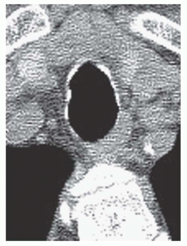
FIG. 22.2. Normal calcification of tracheal cartilage in an elderly woman. Discontinuous calcification of the tracheal wall reflects calcification of individual cartilage rings. The tracheal wall appears thin.

Metastases to the trachea may occur via direct extension or by hematogenous spread. Direct extension to involve the trachea most often is secondary to a primary tumor of the lung, larynx, esophagus, or thyroid. These tumors may compress the trachea, displacing tracheal cartilage inward, or may invade the tracheal lumen, with tumor being seen as abnormal tissue internal to tracheal cartilage (Fig. 22-7; also see Fig. 3-38 in Chapter 3). Hematogenous metastases usually originate from melanoma, or from carcinomas of the breast, colon, or kidney. On CT, hematogenous metastases may appear as single or multiple, sessile or pedunculated endotracheal lesions (Fig. 22-8).

Squamous cell papilloma is the most common benign tracheal tumor. It represents an abnormal proliferation of squamous epithelium, and may appear sessile, papillary, lobulated, or polypoid. Solitary papilloma is associated with smoking and is most common in adults. The condition of multiple papillomas (i.e., papillomatosis) usually begins in childhood with laryngeal involvement and is associated with human papillomavirus infection. On CT, a solitary papilloma appears as a well-circumscribed nodule that is confined to the tracheal wall and projects into the tracheal lumen; it often shows acute angles where it contacts the tracheal wall. Tracheal cartilage is unaffected. Papillomatosis is characterized by numerous nodules involving the entire length of the trachea (see Fig. 3-56 in Chapter 3) or diffuse thickening of the tracheal wall (Fig. 22-9). Other benign tracheal tumors include hamartoma and tumors of mesenchymal origin such as lipoma or chondroma.


FIG. 22.3. Normal expiratory CT. A: On inspiration, the trachea has a rounded appearance. B: During a dynamic forced expiratory scan, there is marked anterior bowing of the posterior tracheal membrane (arrow). This appearance is normal. Little side-to-side narrowing occurs because of the tracheal cartilage.


TABLE 22.1 Tracheal Tumors


Primary malignant


Most common (85% of cases)


Squamous cell carcinoma


Adenoid cystic carcinoma


Rare


Other types of bronchogenic carcinoma


Carcinoid tumor


Sarcoma


Lymphoma


Metastatic


Direct invasion most common


Thyroid carcinoma


Laryngeal cancer


Lung cancer


Esophageal cancer


Hematogenous metastases


Melanoma


Breast carcinoma


Colon carcinoma


Kidney carcinoma


Benign


Squamous cell papilloma


Papillomatosis


Hamartoma


Mesenchymal tumors


FIG. 22.4. Adenoid cystic carcinoma of the proximal trachea. An eccentric narrowing (arrows) of the tracheal lumen is caused by a sessile mass arising from the right tracheal wall.


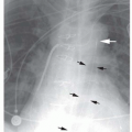
FIG. 22.5. Adenoid cystic carcinoma of the trachea. A: A large mass (large arrows) in the upper trachea markedly narrows the tracheal lumen (small arrow) and invades the mediastinum. B: On a sagittal reconstruction, the mass (arrows) can be seen to arise from the posterior tracheal wall. This location is typical of adenoid cystic carcinoma.


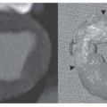
FIG. 22.6. CT appearances of primary tracheal tumor. A: Tracheal malignancies may appear polypoid, sessile, or circumferential. B: Adenoid cystic carcinoma results in a sessile mass (M) arising from the posterior tracheal wall and protruding into the tracheal lumen. The mass extends into the adjacent mediastinum (arrows).


FIG. 22.7. Esophageal carcinoma with tracheal invasion. Tumor has invaded the trachea and is seen as soft tissue (small arrows) internal to the calcified tracheal cartilage (large arrows). The tracheal lumen is narrowed.


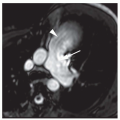
FIG. 22.8. Tracheal metastasis. Tracheal metastases may result in tracheal compression with inward displacement of the tracheal wall, an endotracheal mass, or a combination of these findings. A, B: There is narrowing of the trachea with an endoluminal mass (arrow). C: Soft-tissue window at the same level as (B) shows a mass involving the right tracheal wall and mediastinal soft tissues (arrows).


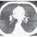
FIG. 22.9. Tracheobronchial papillomatosis with tracheal wall thickening. A: Concentric thickening of the tracheal wall (arrows) is due to diffuse involvement by papillomas. The tracheal lumen is markedly narrowed. B: Multiple cystic pulmonary lesions may be seen in some patients with tracheobronchial papillomatosis.


Tracheomalacia

Tracheomalacia refers to weakness of the tracheal wall, usually due to abnormalities of the tracheal cartilage, associated with excessive collapsibility of the trachea on expiration (Table 22-2). It may be congenital and associated with deficient cartilage, but most often is acquired as a result of intubation injuries, tracheal compression by extrinsic masses or vascular lesions (e.g., aortic aneurysm), chronic infection, chronic obstructive pulmonary disease, or in association
with saber-sheath trachea, relapsing polychondritis, or tracheobronchomegaly. It may be localized, or it may involve a long tracheal segment.


TABLE 22.2 Non-Neoplastic Tracheal Diseases


Disease


Extent


Appearance


Associations


Tracheomalacia


Focal or diffuse


Collapse of tracheal wall with expiration


Congenital intubation, extrinsic masses, chronic infection, COPD, saber sheath trachea, polychondritis, tracheobronchomegaly


Tuberculosis


Distal trachea (and main bronchi)


Early: concentric wall thickening


Late: stricture or distortion of cartilage


Mediastinal lymph node disease


Post intubation stenosis


Proximal trachea


Early: concentric wall thickening


Late: stricture or distortion of cartilage


Malacia may be present


Saber-sheath trachea


Early: thoracic inlet


Late: intrathoracic trachea


Side-to-side narrowing, sagittal diameter normal or increased


COPD, chronic cough; malacia may be present


Wegener’s granulomatosis


Focal (subglottic trachea) or diffuse


Concentric wall thickening, cartilage destruction


Occurs in 15-25% of cases; malacia not often present


Amyloidosis


Focal (nodular) or diffuse


Concentric or nodular wall thickening


Calcification common; malacia is not present


Tracheobronchopathia osteochondroplastica


Diffuse


Calcified submucosal nodules adjacent to tracheal cartilage, nodules spare the posterior membrane


Malacia is not present


Relapsing polychondritis


Diffuse


Thickening of anterior and lateral wall, posterior membrane normal


Arthritis; malacia may be present


Tracheobronchomegaly (Mounier-Kuhn syndrome)


Diffuse


Increased tracheal diameter (>3 cm), tracheal wall normal or appears scalloped, tracheal diverticula


Cystic bronchiectasis often present; COPD, pulmonary fibrosis, Marfan’s syndrome, cystic fibrosis, Ehlers-Danlos syndrome, cutis laxa

Only gold members can continue reading. Log In or Register to continue

Stay updated, free articles. Join our Telegram channel

Oct 10, 2016 | Posted by in CARDIOVASCULAR IMAGING | Comments Off on The Trachea

Full access? Get Clinical Tree

Get Clinical Tree app for offline access