Diffuse Lung Diseases Associated with Calcification


Diffuse Lung Diseases Associated with Calcification


W. Richard Webb


A wide variety of lung diseases may be associated with some degree of lung calcification, most notably granulomatous diseases such as sarcoidosis or tuberculosis (Table 21-1).

Several diffuse lung diseases are associated with extensive lung calcification as a common finding or as a primary manifestation of the disease. These are discussed in this chapter.


DIFFUSE PARENCHYMAL AMYLOIDOSIS

The term amyloidosis refers to a group of conditions characterized by extracellular deposition of abnormal fibrillar protein. Amyloidosis may be classified according to its etiology, the proteins involved, or its primary manifestation (Table 21-2). The first two of these categories overlap, are somewhat confusing, and are reviewed briefly. Classification according to its primary manifestation is most appropriate to the radiologic assessment of amyloidosis and is easiest to remember.


Classification by Etiology

An etiologic classification of amyloidosis includes (1) primary amyloidosis (i.e., associated with a plasma cell disorder or idiopathic); (2) secondary amyloidosis (i.e., associated with chronic inflammatory disorders or neoplasms such as Hodgkin’s disease); (3) heredofamilial amyloidosis (i.e., having a genetic basis); and (4) senile amyloidosis, which usually occurs in patients over the age of 70.

It may be subdivided into systemic forms (i.e., diffuse involvement of multiple organs) or localized forms (i.e., focal involvement largely limited to one organ).


Classification by Specific Proteins

When amyloidosis is classified according to the specific abnormal protein involved, the most important types producing lung disease are amyloid L and amyloid A.

Amyloid L (AL, or light chain amyloidosis) is the most common form of systemic amyloidosis. It may be either idiopathic or associated with plasma cell disorders (about 90% of cases). The lung is involved in 70% to 90% of patients with AL amyloidosis. Cardiomyopathy almost always is present if the lung is involved. Prognosis generally is poor. AL amyloidosis also may result in localized lung involvement. AL typically occurs in patients in their 50s or 60s.

Amyloid A (AA) occurs as secondary amyloidosis in patients with chronic inflammatory diseases (e.g., collagen vascular disease, infections, inflammatory bowel disease, and familial Mediterranean fever) and some neoplasms such as Hodgkin’s disease. The lung is commonly involved to some degree, but deposits often are small or clinically insignificant. The prognosis is better than that for AL.

Among patients who have pulmonary involvement by amyloid, about 65% have primary systemic amyloidosis (systemic AL), 30% have localized amyloidosis (localized AL), and 5% have secondary amyloidosis (AA).


Classification by Manifestation

From a radiographic standpoint, it is most useful to classify amyloidosis on the basis of the abnormalities it produces. In general, pulmonary amyloidosis is classified
into three types: (1) diffuse or alveolar septal amyloidosis, (2) localized nodular amyloidosis, and (3) tracheobronchial amyloidosis.


TABLE 21.1 Diffuse or Multifocal Lung Calcification


Infectious diseases


Tuberculosis


Histoplasmosis


Healed varicella pneumonia


Parasitic infections


Sarcoidosis


Silicosis and coal-worker’s, pneumoconiosis


Calcified metastases


Osteogenic and chondrosarcoma


Mucinous adenocarcinoma


Thyroid carcinoma


Pulmonary ossification with mitral stenosis


Lung fibrosis with dystrophic calcification


Amyloidosis


Alveolar microlithiasis


Metastatic calcification


TABLE 21.2 Classification of Amyloidosis


By etiology


Primary amyloidosis—plasma cell disorder or unassociated with underlying disease


Secondary amyloidosis—associated with chronic inflammatory disorders or neoplasm


Heredofamilial amyloidosis


Senile amyloidosis—usually in patients over 70


Systemic (diffuse involvement of multiple organs)


Localized (focal involvement largely limited to one organ)


By specific proteins


Amyloid L (AL; light-chain amyloidosis)


Most common


Usually primary amyloidosis


Lung involved in 70%-90%, localized or diffuse


Cardiac involvement


Poor prognosis


Amyloid A


Secondary amyloidosis


Lung involved to a minor degree


Better prognosis


By manifestation


Diffuse (alveolar septal) amyloidosis


Usually primary systemic and AL


Dyspnea due to lung or heart disease


Small nodules or reticular opacities


Consolidation


Lymph node enlargement


Calcification common


Localized nodular amyloidosis


Usually primary localized and AL


Usually asymptomatic


Lung nodules, often calcified


Nodules grow slowly


Localized tracheobronchial amyloidosis


Usually primary localized and AL


Tracheal and bronchial narrowing or masses


Calcification common


Diffuse (Alveolar Septal) Amyloidosis

Symptoms of respiratory insufficiency are common with diffuse or alveolar septal amyloidosis, and may be related to diffuse lung disease or associated cardiac involvement. This form of amyloidosis is most often related to primary systemic amyloidosis (systemic AL). Diffuse lung involvement is much less common and may be incidental with AA amyloidosis or senile amyloidosis. Occasionally, diffuse alveolar septal amyloidosis reflects localized rather than systemic disease.

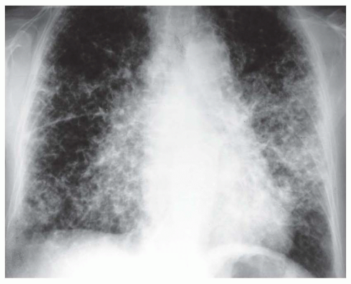
The lung may be diffusely involved in the absence of a radiographic abnormality. If radiographs are abnormal, a reticular or reticulonodular pattern typically is seen, usually bilateral and diffuse or with a basal and subpleural predominance (Figs. 21-1 and 21-2A). Focal or patchy areas of consolidation also may be seen. The abnormal areas can calcify or, rarely, show frank ossification. Less often, a small nodular pattern mimicking sarcoidosis or miliary tuberculosis may be seen.

Hilar or mediastinal lymph node enlargement may be seen in patients with AL, either as an isolated finding or in association with interstitial disease. Cardiac infiltration with heart failure may result in pulmonary edema or pleural effusion.

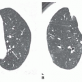
On HRCT, the most common pulmonary manifestations of diffuse parenchymal amyloidosis consist of multiple small nodules, usually ranging from 2 to 4 mm in diameter (see Fig. 21-2B and C), interlobular septal thickening, fine reticular opacities, focal consolidation, foci of calcification within nodules or areas of consolidation (Fig. 21-3), and traction bronchiectasis. A subpleural and basal predominance may be seen.

Progression of the diffuse parenchymal disease typically occurs with time, with an increase in the reticular opacities, septal thickening, the size and number of nodules, the extent of consolidation, and the size and number of calci-fications.

As on chest radiographs, other findings seen on CT include lymphadenopathy, lymph node calcification (Fig. 21-4), and pleural effusion. Lymph node enlargement occurs in up to 75% of cases.


FIG. 21.1. Diffuse alveolar septal amyloidosis. A diffuse reticulonodular pattern is visible, with the abnormality predominating at the lung bases. Lung and lymph node calcification is present, and the heart is enlarged.

Only gold members can continue reading. Log In or Register to continue

Stay updated, free articles. Join our Telegram channel

Oct 10, 2016 | Posted by in CARDIOVASCULAR IMAGING | Comments Off on Diffuse Lung Diseases Associated with Calcification

Full access? Get Clinical Tree

Get Clinical Tree app for offline access