The Idiopathic Interstitial Pneumonias


The Idiopathic Interstitial Pneumonias


Richard W. Web


The idiopathic interstitial pneumonias (IIPs) are a heterogeneous group of diffuse lung diseases occurring without known cause and associated with varying degrees of interstitial lung inflammation and fibrosis. The IIPs have been classified by a consensus committee of the American Thoracic Society and the European Respiratory Society into seven types, based on their histologic pattern, clinical features, and radiographic appearances (Table 13-1). Each of the IIPs is referred to using an acronym. Knowing these acronyms is appropriate, as they are in common usage.

In order of decreasing frequency, the IIPs are as follows:


  • Usual interstitial pneumonia (UIP) and idiopathic pulmonary fibrosis (IPF)


  • Nonspecific interstitial pneumonia (NSIP)


    TABLE 13.1 Idiopathic Interstitial Pneumonias: Classification and Differential Diagnosis


    Histologic Pattern


    Idiopathic Clinical Syndrome


    Differential Diagnosis of the Histologic Pattern


    Usual interstitial pneumonia (UIP)


    Idiopathic pulmonary fibrosis (IPF)


    Collagen-vascular disease, asbestosis, drug toxicity, radiation, chronic hypersensitivity pneumonitis, familial pulmonary fibrosis


    Nonspecific interstitial pneumonia (NSIP)


    Nonspecific interstitial pneumonia (NSIP)


    Collagen-vascular disease, hypersensitivity pneumonitis, drug toxicity, infection, immunodeficiency


    Organizing pneumonia (OP); also known as bronchiolitis obliterans organizing pneumonia (BOOP)


    Cryptogenic organizing pneumonia (COP)


    Infection, bronchial obstruction, aspiration, drug reactions, collagen vascular disease, toxic-fume inhalation, radiation pneumonitis, hypersensitivity pneumonitis


    Diffuse alveolar damage (DAD)


    Acute interstitial pneumonia (AIP)


    Acute respiratory distress syndrome (ARDS) of known cause (e.g., sepsis, shock, infection, drug toxicity, toxic inhalations, trauma)


    Respiratory bronchiolitis (RB)


    Respiratory bronchiolitis-interstitial lung disease (RB-ILD)


    Smoking (usually), inhalations


    Desquamative interstitial pneumonia (DIP)


    Desquamative interstitial pneumonia (DIP)


    Smoking (usually), inhalations


    Lymphoid interstitial pneumonia (LIP)


    Lymphoid interstitial pneumonia (LIP)


    Collagen-vascular diseases (particularly Sjögren’s syndrome, rheumatoid arthritis, and lupus), immunologic disorders (e.g., Hashimoto’s thyroiditis, autoimmune hemolytic anemia, myasthenia gravis, pernicious anemia, chronic active hepatitis), infection, immunodeficiency (e.g., AIDS), drug toxicity


  • Cryptogenic organizing pneumonia (COP)


  • Acute interstitial pneumonia (AIP)


  • Respiratory bronchiolitis-interstitial lung disease (RB-ILD)


  • Desquamative interstitial pneumonia (DIP)


  • Lymphoid interstitial pneumonia (LIP)

In this classification, a distinction is made between the histologic pattern of the IIP and the idiopathic clinical syndrome associated with it. In several cases, these have different names (see Table 13-1).

Although the IIPs are designated as idiopathic, identical histologic patterns, radiographic appearances, and clinical symptoms may occur in association with specific diseases or exposures, including collagen-vascular disease, drug reactions, smoking, and infection (see Table 13-1). It is
helpful to think of the histologic patterns seen in the IIP as having a differential diagnosis, with one of the possible causes being an idiopathic syndrome (see Table 13-1). Since the radiographic appearances of the IIP parallel their histologic patterns, this approach allows the appropriate radiographic or high- resolution CT (HRCT) differential diagnosis to be suggested.


USUAL INTERSTITIAL PNEUMONIA AND IDIOPATHIC PULMONARY FIBROSIS

IPF is the idiopathic disease associated with the histologic pattern termed usual interstitial pneumonia.

Histologically, UIP is characterized by a heterogeneous pattern with different parts of the biopsy showing normal lung, interstitial inflammation, fibroblastic proliferation, interstitial fibrosis, and honeycombing; in other words, the biopsy shows spatial heterogeneity. Histologic abnormalities appear to represent different stages in the temporal evolution of fibrosis, a combination of end-stage and more active lesions. This is termed temporal heterogeneity. Spatial and temporal heterogeneity is characteristic of UIP and is not seen in other IIPs. Lung involvement is patchy, although there is a predominance in the subpleural and basal lung regions (Table 13-2).

UIP may be associated with collagen-vascular disease, asbestosis, drug toxicity, radiation, chronic hypersensitivity pneumonitis, or familial pulmonary fibrosis. Idiopathic UIP is termed idiopathic pulmonary fibrosis. IPF accounts for about 70% of cases of UIP.

IPF occurs most frequently in patients older than 50 (see Table 13-2). Symptoms include progressive dyspnea, cough, weight loss, and finger clubbing; symptoms usually precede presentation by 6 months. Pulmonary function tests (PFTs) show restriction with a reduced diffusing capacity. Treatment is largely ineffective. IPF has a poor prognosis, with a mean survival of 3 to 4 years from the onset of symptoms. The 5-year survival rate is 25% to 40%. Lung transplantation may be required.

The diagnosis of IPF is limited to patients with histologic findings of UIP; in other words, if it is not UIP, it is not IPF. Histologic diagnosis requires an open lung biopsy; transbronchoscopic biopsy is not sufficient for diagnosis but may be used to exclude other causes of lung disease.

Based on a 2002 consensus of the American Thoracic Society and European Respiratory Society, a diagnosis of IPF in the absence of lung biopsy can be based on a combination of clinical, functional, and HRCT findings. Based on this consensus, IPF is considered to be likely if all four major criteria and three of four minor criteria are present (Table 13-3). These criteria outline common features of this disease.

In a 2010 revision of these criteria, the diagnosis of IPF has been simplified to a combination of


TABLE 13.2 Usual Interstitial Pneumonia (UIP) and Idiopathic Pulmonary Fibrosis (IPF): Histologic, Clinical, and HRCT Features


UIP: histologic pattern


Characteristics


Spatial and Temporal inhomogeneity


Dense fibrosis, often with honeycombing


Fibroblastic foci


Patchy lung involvement


Subpleural and basal predominance


Differential diagnosis


Idiopathic (IPF)


Collagen-vascular disease


Asbestosis


Drug toxicity


Radiation


Chronic hypersensitivity pneumonitis


Familial pulmonary fibrosis


IPF: idiopathic clinical syndrome


Age <50 years


Progressive dyspnea; duration of symptoms 3 months or more


Velcro rales


Pulmonary function tests: restriction and impaired gas exchange


Treatment largely ineffective


5-year survival rate 25%-40%


HRCT findings highly accurate in diagnosis


Honeycombing


Reticulation


Subpleural and basal predominance


Little ground-glass opacity


  • A UIP pattern on HRCT or histopathology


  • The absence of an alternative etiology (e.g., collagen-vascular disease, asbestos exposure, drug treatment, hypersensitivity pneumonitis) after careful clinical evaluation (i.e., history, physical examination, pulmonary function testing, and laboratory assessment)


TABLE 13.3 Criteria for a Clinical Diagnosis of Idiopathic Pulmonary Fibrosis (2002)


Major criteria


Exclusion of known causes of infiltrative lung disease such as exposures, drugs, and connective tissue disease


Abnormal pulmonary function tests with evidence of restriction and impaired gas exchange


HRCT findings of bibasilar reticulation with minimal ground-glass opacity


Transbronchial lung biopsy or bronchoalveolar lavage showing no evidence of another disease


Minor criteria


Age <50 years


Insidious onset of dyspnea on exertion


Duration of illness of ≤3 months


Bibasilar inspiratory crackles (so-called Velcro rales)


Radiographic Findings

In 80% of patients with UIP/IPF, radiographs show a bilateral reticular pattern predominantly involving the lower lung zones and subpleural lung regions (Figs. 13-1 and 13-2). In its earliest stages, a fine reticular pattern may be visible in the posterior costophrenic angles on the lateral radiograph (see Fig. 13-1C and 13-2B), and the lateral radiograph is often more sensitive than the PA view in making this diagnosis. As fibrosis progresses, the reticular pattern becomes coarse, and cystic areas of honeycombing are visible in about half of cases (see Fig. 13-2). Pleural thickening or effusion is very uncommon.


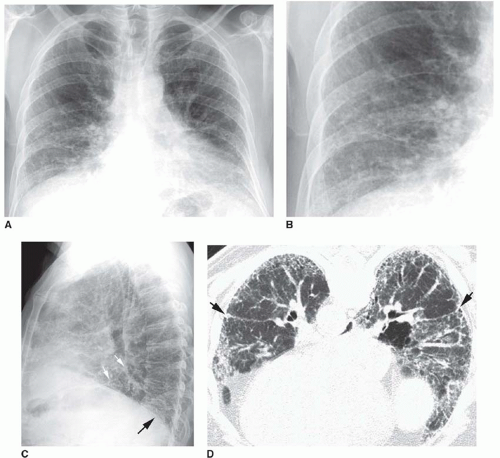
FIG. 13.1. Chest radiograph and HRCT in a patient with histologically proven idiopathic pulmonary fibrosis. A: PA radiograph shows reduced lung volumes. There is an increase in reticular opacities in the lung periphery and at the lung bases. The appearance and distribution are typical of idiopathic pulmonary fibrosis. B: Detail view of the right lower lobe shows increased reticular opacities. C: Lateral view shows increased reticular opacities in the posterior costophrenic angles (black arrow). A major fissure (white arrows) is bowed posteriorly because of more severe fibrosis in the lower lobe. D: Prone HRCT shows extensive subpleural reticular opacities with mild honeycombing. The major fissures (arrows) are displaced posteriorly because of lower lobe fibrosis.

Because of lung fibrosis, decreased lung volume is typical (see Fig. 13-1A), and serial chest radiographs are often used to monitor progressive reduction in lung volume with time. Because of greater fibrosis and volume loss in the lower lobes, the major fissures may be seen to bow posteriorly (see Fig. 13-1C).


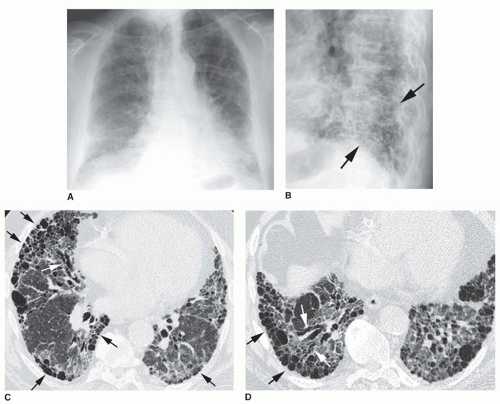
FIG. 13.2. Chest radiograph and HRCT in a patient with idiopathic pulmonary fibrosis. A: PA radiograph shows ill-defined reticular opacities at the lung bases. B: Coned-down lateral view shows a coarse reticular pattern (arrows) in the posterior costophrenic angles. C and D: HRCT shows extensive subpleural honeycombing (black arrows), reticular opacities, and traction bronchiectasis (white arrows). The honeycomb cysts range from a few millimeters to 2 cm in diameter.


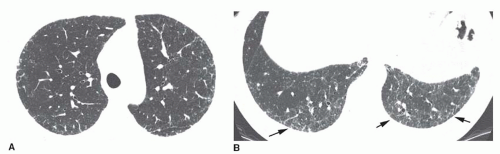
FIG. 13.3. Reticulation in early usual interstitial pneumonia/idiopathic pulmonary fibrosis (UIP/IPF). A and B: Concentric subpleural reticulation is the only abnormality. It is most severe in the lung bases (arrows B). Reticulation is the earliest abnormality seen on HRCT in patients with IPF. This appearance is nonspecific. Biopsy would be necessary for diagnosis.


A combination of clinical and radiographic findings is sufficient to make an accurate diagnosis of IPF in about 70% of cases. In 10% of cases, the radiograph appears normal.


HRCT Findings

On HRCT, UIP/IPF is usually characterized by the presence of honeycombing, irregular reticular opacities, and traction bronchiectasis (see Figs. 13-1 and 13-2 and Table 13-2). In its earliest stages, the only HRCT abnormality may be fine reticulation in the subpleural lung (Fig. 13-3); as the disease progresses, the reticular opacities coarsen and become more extensive, and traction bronchiectasis develops. Honeycombing is first seen in the immediate subpleural lung and is present in about 70% of cases of IPF (Figs. 13-1D, 13-2C and D, 13-4, 13-5). In severe cases, findings of honeycombing predominate (Fig. 13-5). Honeycomb cysts usually range from 2 mm to 2 cm in diameter but may be larger.

On HRCT, findings of UIP/IPF typically predominate in the peripheral, subpleural regions and in the lung bases. A subpleural predominance is evident on HRCT in 80% to 95%, and either concentric (Fig. 13-5) or patchy (Fig. 13-6) subpleural honeycombing is characteristic. In approximately 80% of patients, the fibrosis is most severe in the lower lung zones; in about 20%, all zones are involved to a similar degree. If the upper lobes appear abnormal, the extent and severity of abnormalities are typically less than at the lung bases. Subpleural, basal honeycombing associated with irregular reticular opacities in the upper lobes is characteristic of IPF. Findings of fibrosis and honeycombing may be asymmetrical (Fig. 13-7).


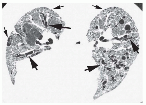
FIG. 13.4. Reticular opacities, traction bronchiectasis, and honeycombing in idiopathic pulmonary fibrosis. Prone HRCT shows a diffuse, coarse, reticular pattern at the lung bases, with traction bronchiectasis (large arrows) and mild subpleural honeycombing (small arrows).


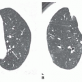
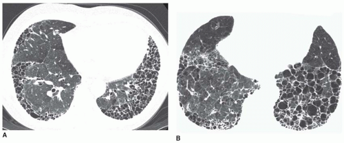
FIG. 13.5. Honeycombing on HRCT in two patients with idiopathic pulmonary fibrosis. A and B: Concentric subpleural honeycombing is visible in the lung bases. Most cysts are less than 2 cm in size.

Ground-glass opacity may be seen in patients with UIP/IPF, but this finding typically reflects the presence of fine lung fibrosis. Ground-glass opacity is almost always seen in association with reticulation and traction bronchiectasis in the same lung regions (Fig. 13-8); it is rare as an isolated finding.

Mediastinal lymph node enlargement is visible on CT in more than 70% of cases. The enlarged nodes typically measure less than 15 mm.

A confident HRCT diagnosis of UIP may be based on the presence of reticular opacities associated with honeycombing and/or traction bronchiectasis, with a predominant basal and
subpleural distribution. If these findings are present, the accuracy of HRCT in making the diagnosis of UIP exceeds 90%. In the absence of an associated disease (e.g., collagen-vascular disease) or exposure (e.g., asbestos, drugs, organic antigens), IPF is very likely the diagnosis. In patients with typical HRCT and clinical findings, lung biopsy is uncommonly performed.


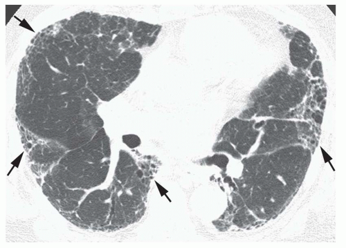
FIG. 13.6. Patchy subpleural reticular opacities and honeycombinga in idiopathic pulmonary fibrosis. Areas of honeycombing (arrows) involve the peripheral lung in a patchy fashion.

In the vast majority of patients with IPF, serial HRCT scans show an increase in the extent of reticulation and honeycombing. This progression usually occurs gradually over several months or years. Occasionally, a patient develops a fulminant and sometimes fatal acute exacerbation characterized by consolidation or ground-glass opacity (Fig. 13-9); biopsy typically shows diffuse alveolar damage (DAD). When such exacerbations occur, pulmonary function decreases in a series of acute stair-step increments coincident with the exacerbations.


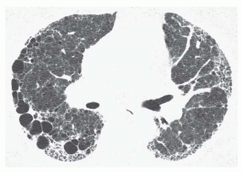
FIG. 13.7. Asymmetrical honeycombing in idiopathic pulmonary fibrosis. Subpleural reticulation and honeycombing are present. The honeycomb cysts are much larger on the right.


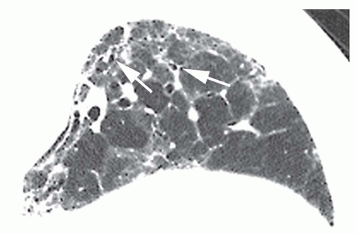
FIG. 13.8. Ground-glass opacity in idiopathic pulmonary fibrosis. Detail of the left lower lobe on a prone HRCT shows patchy ground-glass opacity. These regions also show irregular reticular opacities and traction bronchiectasis (arrows) and bronchiolectasis, indicating the presence of fibrosis. Ground-glass opacity is rare in idiopathic pulmonary fibrosis as an isolated finding but, as in this case, may be seen in association with reticular opacities.


NONSPECIFIC INTERSTITIAL PNEUMONIA

NSIP is less common than UIP. The histologic pattern and the IIP are both called NSIP.

Despite its name, it is best to think of NSIP as a specific pathologic entity, not a “nonspecific” diagnosis. NSIP is characterized histologically by alveolar wall thickening or fibrosis. Abnormalities are described as showing spatial and temporal homogeneity. In other words, all parts of the biopsy look the same and represent the same stage of the disease; in UIP/IPF, the biopsy appears heterogeneous. NSIP lacks specific features that would allow a diagnosis of another interstitial pneumonia. It may demonstrate cellular (i.e., cellular NSIP) or fibrotic (i.e., fibrotic NSIP) patterns (Table 13-4).

NSIP may be idiopathic or may be associated with collagen-vascular diseases, hypersensitivity pneumonitis, drug toxicity, infection, or immunodeficiency. An association with collagen-vascular diseases is particularly common, and NSIP is the most common histologic abnormality present in patients with collagen disease and a lung abnormality.

Clinically, patients with NSIP present with symptoms similar to those of IPF, including dyspnea and cough. The average duration of symptoms is of 18 to 30 months at diagnosis (see Table 13-4). Average age at diagnosis is 40 to 50 years. Patients typically respond to treatment with steroids and the prognosis is good, although this varies with the degree of fibrosis present. Overall, 5-year survival rate is 80% to 90%. Patients with cellular NSIP have a 5-year survival rate exceeding 90%; in patients with fibrotic NSIP, 5-year survival
rate is 65% to 90%. Patients with NSIP may suffer an acute exacerbation similar to those occurring in UIP/IPF.


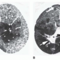
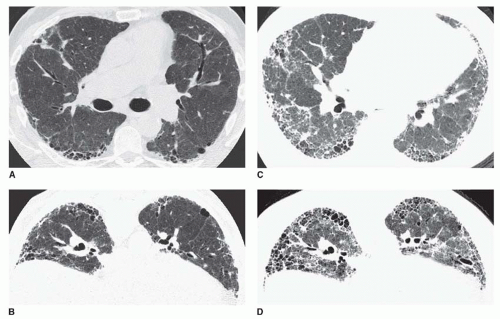
FIG. 13.9. Acute exacerbation of idiopathic pulmonary fibrosis. Supine (A) and prone (B) HRCTs show subpleural reticulation and honeycombing in the lower lobes, typical of idiopathic pulmonary fibrosis. Supine (C) and prone (D) HRCTs at the time of an acute exacerbation show progression of honeycombing and an increase in ground-glass opacity. This reflects diffuse alveolar damage.


Radiographic Findings

The radiographic findings consist mainly of ill-defined opacity, ground-glass opacity, or consolidation predominantly involving the lower lung zones (Figs. 13-10A and 13-11A). Other manifestations include a reticular pattern or a combination of reticular and air-space patterns. In 10% or more of cases, the chest radiograph is normal.


HRCT Findings

Because the histology associated with NSIP is variable, HRCT may show a variable combination of ground-glass opacity, patchy consolidation, irregular reticular opacities, and traction bronchiectasis, usually with a peripheral and basal predominance. Although honeycombing may be present in patients with NSIP, it is uncommon, being seen in a few percent of cases, and when present, tends to be inconspicuous and limited in extent, particularly in comparison to patients with UIP and IPF.

In most cases, patchy or concentric subpleural ground-glass opacity with a lower lobe predominance is seen on HRCT; superimposed reticulation is common, being seen in 50% of cases (see Figs. 13-10,13-11,13-12 and 13-13). Although it predominates in the peripheral lung, NSIP may show sparing of the immediate subpleural region (20% to 50% of cases), a finding that strongly suggests this diagnosis and helps distinguish NSIP from UIP and IPF (see Figs. 13-12,13-13,13-14,13-15 and 13-16). The presence of traction bronchiectasis often indicates fibrotic NSIP (Figs. 13-15 and 13-16), but also may be seen in cellular NSIP. Reticulation, traction bronchiectasis, and honeycombing occurring in the absence of ground-glass opacity correlate with the fibrotic pattern of NSIP. The cellular pattern of NSIP typically results in ground-glass opacity (Fig. 13-12), sometimes combined with reticulation or traction bronchiectasis; ground-glass opacity may resolve with treatment (Fig. 13-17). Abnormalities may be quite subtle in early or mild cases, being visible with certainty only on prone scans.

Only gold members can continue reading. Log In or Register to continue

Stay updated, free articles. Join our Telegram channel

Oct 10, 2016 | Posted by in CARDIOVASCULAR IMAGING | Comments Off on The Idiopathic Interstitial Pneumonias

Full access? Get Clinical Tree

Get Clinical Tree app for offline access