51 Continuous Moving-Table Imaging
Continuous moving-table imaging enables large field-of-view, as well as whole-body, imaging to be completed in a single setting. Thus multiple organ systems or anatomic areas can be evaluated efficiently in a short time. Continuous moving-table imaging is particularly useful for ultrashort bore scanners, as it overrides any intrinsic limitation in field-of-view by sliding the patient through the magnet and always acquiring images at the center of the magnet (the isocenter). This type of data acquisition takes advantage of the optimal magnetic field homogeneity and gradient linearity at the center of the magnet.
Continuous scanning during automatic table movement has expanded the applications of MR imaging and opened doors to new possibilities. Any extended anatomic coverage can be selected. Scanning takes place automatically and covers all body parts, with electronically controlled slow movement of the patient through the magnet. Image acquisition occurs continuously as the region of interest slides smoothly through the center of the magnet.
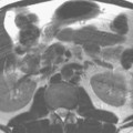
Continuous moving-table imaging can be applied in combination with various techniques including (1) single slice sequential as well as multislice acquisitions, (2) 2D as well as 3D acquisitions, and (3) anatomic as well as vascular acquisitions. Figure 51.1 presents an example of extended anatomic coverage using sequential 2D imaging with T1- and T2-weighted scans, in addition to STIR (courtesy of UCLA). Figure 51.2
Stay updated, free articles. Join our Telegram channel

Full access? Get Clinical Tree


