46 Contrast-Enhanced MRA: Basics; Renal, Abdomen
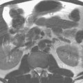
The contrast-enhanced MRA maximum intensity projection (MIP) image displayed in Fig. 46.1A demonstrates the abdominal aorta and common iliac arteries, with moderate to severe stenosis noted at the origin of the left renal artery (arrow). Figure 46.1B demonstrates extensive atherosclerotic disease involving the aorta, with severe stenosis at the origin of the left renal artery. Both studies were performed at 1.5 T.
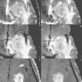
The study presented in Fig. 46.2 (reprinted with permission from U. Kramer, Invest Radiol 2007;42:747) illustrates the feasibility of high spatial resolution contrast-enhanced MRA at 3 T, providing a further improvement in evaluation of the renal artery and its branches. Early branching is demonstrated, involving both renal arteries, in this potential, living, related kidney donor. The voxel size was 1 mm3
Related posts:

Stay updated, free articles. Join our Telegram channel

Full access? Get Clinical Tree


