86 Contrast Media: Gd Chelates with Extracellular Distribution
The gadolinium (Gd) chelates are the dominant class of contrast media for MRI. They are clear, colorless fluids, formulated without bacteriostatic additives for intravenous administration. The standard dose (excluding use in MR angiography) is 0.1 mmol/kg, which corresponds to 15 mL for a 75-kg patient (with all agents except one formulated at a concentration of 0.5 molar). The distribution of the agents is to the extracellular space.
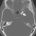
Lesion enhancement occurs by one of two mechanisms: disruption of the bloodbrain barrier (for intraaxial brain lesions) and lesion vascularity. The gadolinium ion is strongly paramagnetic, leading to a reduction in both T1 and T2, which is visualized on T1-weighted images as an increase in signal intensity. In Figure 86.1, thin-section T1-weighted images are illustrated at the level of the internal auditory canal, revealing a soft tissue mass (a vestibular schwannoma) on the right precontrast (Fig. 86.1A), which demonstrates prominent enhancement postcontrast (white arrow, Fig. 86.1B). Enhancement of normal vascular structures includes the nasal turbinates (black arrow, Fig. 86.1B
Stay updated, free articles. Join our Telegram channel

Full access? Get Clinical Tree


