Key Points
- ▪
Anomalies of coronary ostia and course are more common than appreciated.
- ▪
Cardiac CT angiography is the preferred test to characterize anomalies of the coronary ostia and course. The ability to anatomically depict both the structure and course of the coronary artery and the immediately adjacent structures provides comprehensive assessment of coronary anomalies.
- ▪
Cardiac CT is able to provide more extensive (distal) characterization of coronary anomalies than can cardiac MRI.
A wide range of coronary artery anomalies have been described, reflecting the many permutations of abnormalities of the following anatomic details:
- □
The location of the ostium:
- •
Which sinus of Valsalva
- •
Where in the sinus of Valsalva
- •
Ascending aorta
- •
The aortic arch
- •
The descending aorta
- •
The innominate artery
- •
The common carotid artery
- •
The internal thoracic artery
- •
The pulmonary artery
- •
A bronchial artery
- •
The left ventricle
- •
- □
Common versus separate ostium with other coronary arteries
- □
The shape of the ostia (slit-like or not)
- □
Congenital ostial stenosis
- □
Congenital ostial atresia
- □
The initial course of the artery—within the wall of the aorta (i.e., intramural) or not
- □
The angulation of the initial course—tangential or not
- □
The ongoing course of the artery:
- •
Anterior to the pulmonary artery
- •
Intra-arterial (between the aorta and the pulmonary artery)
- •
Through the crista supraventricularis portion of the septum
- •
Dorsal/retroaortic (posterior to the aorta)
- •
- □
Epicardial or intramyocardial course
Understandably, many classifications have been proposed to describe anomalies of the coronary ostia and their course, and the terminology they use often varies. For example, most series would describe “separate ostia of the LCX and LAD,” whereas some describe an “absent left main stem.” Many series and classifications are very detailed, whereas others, particularly the smaller series, are less detailed. Merging the data from different series to arrive at an overall picture is a challenge.
Anomalies of the coronary ostia and their course are the most common anomalies observed at angiography, accounting for 90% of such cases. The remaining 10% are anomalies of termination—i.e., coronary fistulae. Anomalies of the coronary ostia and their course most commonly involve the left coronary artery, especially the left circumflex coronary artery, which accounts for about 60% of observed anomalies of origin and course.
The Most Common Coronary Anomalies
- □
Left circumflex artery (LCX) arising from:
- •
A separate ostia in the right sinus of Valsalva (69%)
- •
LCX arising from the proximal right coronary artery (RCA; 31%)
- •
- □
Single coronary artery from the left sinus of Valsalva
- □
Both coronary arteries arising from the right sinus of Valsalva
- □
Left anterior descending artery (LAD) arising from the right sinus of Valsalva
Overall, the most common course of an anomalous artery is anterior or posterior to the great vessels, rather than intra-arterial, although nearly half of anomalous right coronary arteries have an intra-arterial course.
Terminology of Anomalies of Course
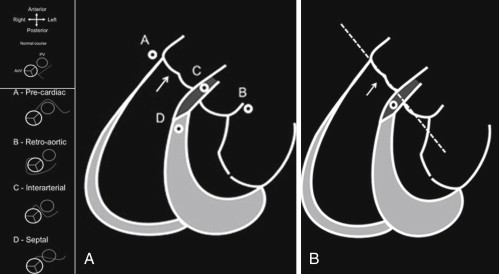
Anomalies of course may be described using the following terminology :
- □
Type A: A nterior to the pulmonary artery, or pre-pulmonic course
- □
Type B: B etween the aorta and pulmonary artery, or intra-arterial course
- □
Type C: Through the c rista supraventricularis; also known as intraseptal, septal, subpulmonary course, or “tunneled”
- □
Through the right ventricular (RV) infundubulum
- □
Type D: D orsal pathway, or posterior” or retro-aortic course
- □
Mixed
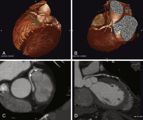
Torres et al. reported on 6000 consecutive cardiac CT (CCT) cases. Of 15 anomalous left coronary arteries arising from the right sinus of Valsalva and anomalous right coronary arteries arising from the left sinus of Valsalva, and coursing between the aorta and the pulmonary artery, the following patterns were seen: two were intra-arterial, four were intraseptal, eight had a mixed intra-arterial/intraseptal course, and one coursed through the right ventricular infundibulum. These findings challenge the traditional classification of the course of anomalous coronary arteries ( Fig. 9-1 ).
Incidence of Coronary Artery Anomalies
The true incidence of coronary anomalies is unknown; furthermore, despite a considerable number of papers describing coronary anomalies in depth, the true range of anomalies is incompletely mapped out. The reported incidence varies depending on the definition, the methodology used, and the patient profile (the pretest probability) assessed.
Incidence by Modality
- □
Echocardiography: 0.1%
- □
Angiography : 1.0%
- □
Autopsy: 0.12%
Incidence by Patient Profile
- □
Asymptomatic individuals: unknown
- □
High school athletes with sudden death: 11%
- □
Competitive athletes (<35 years) with sudden death : 13%
- □
Military recruits with nontraumatic sudden death : 33%
- □
Angiography/bypass surgery (CAD) Coronary Artery Surgery Study (CASS): 0.3%
Determining the Clinical Relevance of Coronary Artery Anomalies
Most patients with coronary anomalies are asymptomatic throughout life, and most coronary anomaly patterns are believed to be benign (∼70–80%). Only about 20% to 30% of those recognized are considered potentially serious or lethal.
Coronary anomalies that do not commonly have clinical risk include those with high ostia, multiple ostia, or “split” courses, and those that do not plausibly impair coronary flow (i.e., do not have congenital stenosis or atresia, do not have a tangential take-off, do not have an intramural course, do not have an intra-arterial course) or supply with desaturated blood (i.e., do not arise from the pulmonary artery).
Establishing that a coronary anomaly is the cause of clinical symptoms or presentation requires that alternative explanations and plausible associations be excluded.
Occasional clinical manifestations include sudden death, ventricular fibrillation, myocardial infarction, cardiomyopathy, syncope, and chest pain.
Major clinical presentations appear to be more common with certain patterns of anomalies and with certain patient groups. Among high school athletes who experience sudden nontraumatic death, coronary anomalies are the second most common cause of death. Similarly, it has been reported that coronary artery anomalies may be the second most common cause of sudden death in competitive athletes less than 35 years of age (second only to hypertrophic cardiomyopathy). , In an autopsy series of military recruits, coronary artery anomalies were the most common cause of sudden nontraumatic death.
A given anomaly, though, may have disparate clinical profiles: it may be found in a young patient who is symptomatic and who is being assessed for the these symptoms, or it may be “incidentally” detected well into adult life when angiography for coronary artery disease (CAD) evaluation or CT scanning for other purposes is performed. Many notable cases have been documented where, for example, longevity was achieved by a patient who had an anomaly generally thought to be “potentially serious” or “lethal.”
The many permutations of anomalies lessen their overall clinical relevance; however, several recurrent themes have emerged. Among young athletes with anomalous coronary arteries who experience sudden death, over three quarters of the anomalies have an intra-arterial course. The intra-arterial course of the left main coronary artery (LMCA) is associated with higher risk than a retro-aortic course, although the retro-aortic course may be associated with risk. Although the classic intra-arterial anomaly associated with increased clinical risk is the intra-arterial LMCA, intra-arterial LAD and RCA anomalies also are associated with increased risk.
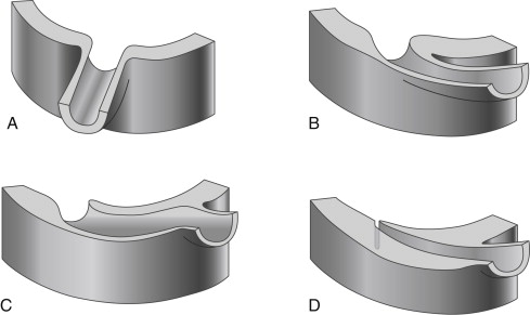
An “intramural” course ( Fig. 9-2 ), completely within the wall of the aorta, appears to be a serious anomaly, associated with sudden death during athletic exertion. An anomalous intramural course often is associated with an angulated initial course. Anatomically, the initial segment of the coronary artery is entirely within the wall of the aorta, and there is one adventitia. Identification of an intramural course requires high-resolution imaging or surgical inspection.
Initially, the intra-arterial course of an anomaly itself was thought to be responsible for the clinical risk, with hypotheses such as compression of the anomalous course by the pulmonary artery trunk and aorta during dilation associated with exercise resulting in myocardial ischemia and clinical risk. It is increasingly being recognized, however, that it is the abnormal ostium or initial course of the coronary artery associated with the intra-arterial course that confers the risk. Abrupt (“tangential”) angulation of the initial course (take-off) may be associated with “kinking” of the artery and a slit-like ostium. It also has been hypothesized that spasm of the anomalous intra-arterial coronary artery contributes to myocardial ischemia and clinical risk.
At the same time, the surgical approach to intra-arterial coronary artery anomalies has evolved beyond aortocoronary bypass grafting to an increased focus on “un-roofing” or “marsupializing” the narrowed ostium/initial course.
Associations of Coronary Anomalies
Most coronary anomalies occur in isolation, i.e., without associated congenital cardiac or vascular anomalies. However, some forms of congenital heart disease are commonly associated with coronary artery anomalies.
Tetralogy of Fallot
Tetralogy of Fallot (ToF) is the prototypical disorder of associated congenital heart disease and coronary anomalies. The anomalies are critically relevant to corrective surgery for ToF, because the coronary artery anomalies render the coronary circulation vulnerable to damage by surgery. The incidence of associated coronary anomalies is 1% to 10%. The most common associated anomalies are:
- □
A large conus artery
- □
An LAD with an anomalous course (LAD arising from the proximal RCA or off the right sinus of Valsalva) that crosses the right ventricular outflow tract (RVOT; 37%). This is more common when the aortic root is more anterior, rightward, or lateral.
- □
A single coronary artery
Delineation of the origin and course of coronary arteries should be achieved before any intervention on the RVOT. CCT is able to detect LAD anomalies associated with ToF.
Transposition of the Great Arteries (TGA)
Transposition of the great arteries (TGA) is often associated with coronary artery anomalies that confer risk of injury to the coronary circulation at the time of surgery. The most common associated anomalies are:
- □
Anomalous origin of the RCA from the posterior right sinus or off the left main from the posterior left sinus, seen in 60% of cases
- □
Anomalous origin of the LCX off the RCA, seen in 16% to 20% of cases
- □
More complex anomalies also seen with TGA.
- •
The LCX arises from the RCA
- •
The LAD arises from the RCA
- •
The LCA arises from the right sinus
- •
Solitary coronary arteries
- •
Intramyocardial courses
- •
- □
Following successful arterial switch operation, intrinsic thickening and extrinsic compression or torsion may occur.
Truncus Arteriosus
Truncus arteriosus infrequently may be associated with coronary anomalies.
Anomalies of Coronary Ostia and Course
See Box 9-1 and Figures 9-3 through 9-7 .
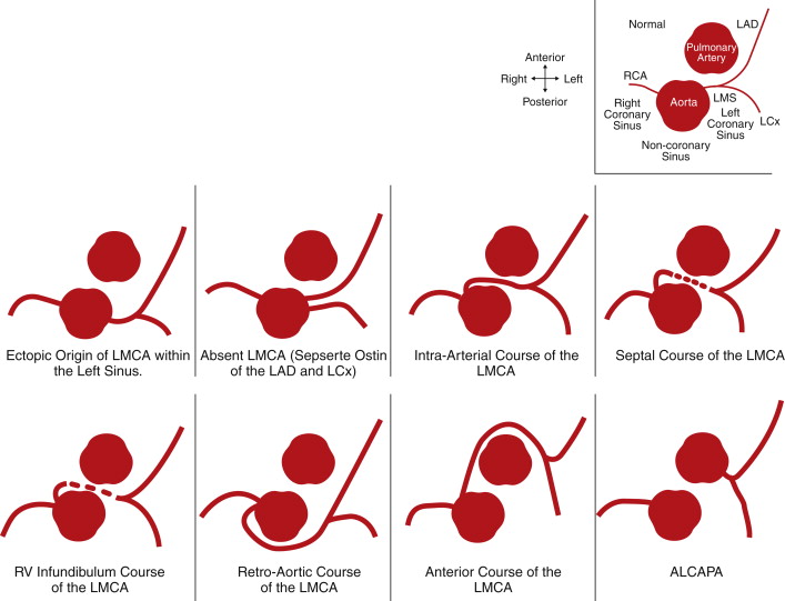
LMCA
High origin (above STJ)
CCT incidence: 0.20%
Angiography incidence: 0.13%
Ectopic origin within the LSV ( Fig. 9-3 )
CCT incidence: 0.59%
Angiography incidence: 0.41%
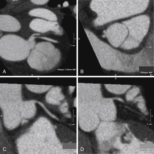
Absent = separate LAD and LCx ostia ( Figs. 9-3, 9-8 )
CCT incidence: 0.59%
Angiography incidence: 0.02%
RCS : arising from a separate ostium, a common ostium, or a common solitary coronary artery with any of the following courses:
Intra-arterial (± a slitlike ostium, ± an intramural course) ( Figs. 9-3, 9-9, 9-11 )
RV infundibuum ( Figs. 9-3, 9-12, 9-13 )
Septal ( Figs. 9-3, 9-14 )
Retroaortic ( Figs. 9-3, 9-15 )
Anterior to the RVOT ( Fig. 9-3 )
Mixed
NCS
Arising from the PA (ALCAPA / BWG syndrome) ( Figs. 9-3, 9-16 to 9-19 ; )
Angiography incidence: 0.01%
Arising from the innominate artery
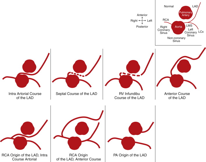
LAD
High origin (above the STJ)
Separate ostium from the LCx ( Fig. 9-3 )
Separate ostium from the LCx abnormal location within the LSV
RCS : arising from a separate ostium, a common ostium, or a common solitary coronary artery with any of the following courses:
Intra-arterial (± a slitlike ostium, ± an intramural course) ( Figs. 9-4, 9-20, 9-21 )
RV infundibuum ( Fig. 9-4 )
Septal ( Fig. 9-4 )
Anterior to the RVOT ( Fig. 9-4 )
RCS : arising from the RCA (may run with an intra-arterial, septal, RV infundibular, retroaortic, or anterior course) ( Fig. 9-4 )
Split LAD: first half from the LMCA continuation and the second half from a large AM branch ( Fig. 9-4 )
Arising from the PA ( Fig. 9-4 )
Dual/bifid LAD:
Type 1: early bifurcation into a short (terminating high within the anterior interventricular groove) and a long LAD parallel to the AIV groove on the left side
Type 2: early bifurcation into a short (terminating high within the anterior interventricular groove) and a long LAD parallel to the AIV groove on the right side
Type 3: early bifurcation into a short (terminating high within the anterior interventricular groove) and a long LAD with an intramyocardial course
Type 4: early bifurcation into a short (terminating high within the anterior interventricular groove) and a long LAD arising from the RCA Intramyocardial courses (“myocardial bridging”)
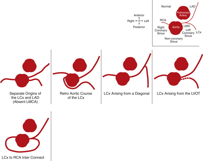
LCx
Separate ostium from the LAD
Separate ostium from the LCx abnormal location within the LSV
RCS: arising from a separate ostium, a common ostium, or a common solitary coronary artery with any of the following courses:
Intra-arterial (± a slitlike ostium, ± an intramural course)
Septal
Retroaortic ( Figs. 9-22, 9-23 ; )
Anterior to the RVOT
Mixed
RCS: arising frm the RCA (may run with an intra-arterial, septal, RV infundibular, retroaortic, or anterior course)
Arising from the LVOT/subaortic
Arising from a diagonal branch
CCT incidence: 0.20%
LCx: RCA interconnection
Angiography incidence: 0.00%
Intramyocardial courses (“myocardial bridging”)
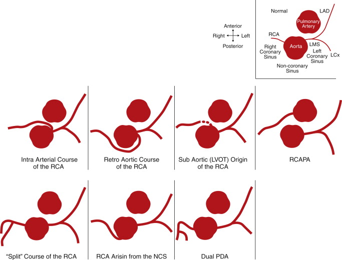
RCA
High origin (above STJ)
Angiography incidence: 0.15%
Ectopic origin within the RSV
CCT incidence: 0.79%
LCS: arising from a separate ostium, a common ostium, or a common solitary coronary artery with any of the following courses:
Intra-arterial (± a slitlike ostium, ± an intramural course) ( Figs. 9-24 to 09-33 ; )
Retroaortic
Mixed
Arising from the LVOT / subaortic
Arising from the PA (RCAPA) 15 ( Fig. 9-34 )
Angiography incidence: 0.00%
Split RCA (first half of the PDA from the RCA and the second half from an AM)
CCT incidence: 0.98%
Arising from the innominate artery ( Fig. 9-35 )
Intra-atrial course
Superdominant RCA (no LCx)
Arising from the NCS
RCA Tunnel
Intramyocardial courses (“myocardial bridging”)
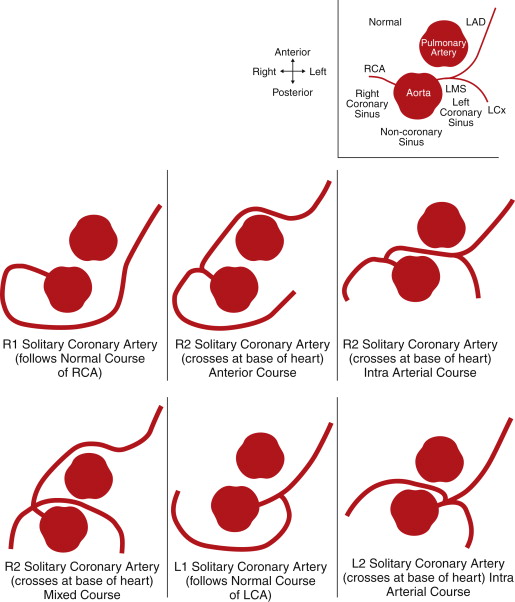
SINGLE CORONARY ARTERY
Arising from the RSV:
R1: Follows the normal course of an RCA ( Fig. 9-36 )
R2: LCA crosses at base of heart ( Figs. 9-37, 9-38 )
R3: LAD and LCx arise separately/absent LMCA
Arsing from the LSV:
L1: Follows the normal course of an LCA
L2: RCA crosses at base of heart
Arising from the NCS
AIV, anterior interventricular vein; ALCAPA, anomalous left coronary artery from the pulmonary artery; AM, acute marginal; CCT, cardiac CT; LAD, left anterior descending; LCA, left coronary artery; LCS, left coronary sinus; LCX, left circumflex coronary artery; LMCA, left main coronary artery; LSV, left sinus of valsalva; LVOT, left ventricular outflow tract; NCS, noncoronary sinus; PA, pulmonary artery; PDA, posterior descending coronary artery; RCA, right coronary artery; RCAPA, right coronary artery arising from the pulmonary artery; RCS, right coronary sinus; RSV, right sinus of valsalva; RV, right ventricle; RVOT, right ventricular outflow tract; STJ, sinotubular junction.
Solitary Coronary Artery
A common ostium to the left and right coronary arteries (i.e., a solitary coronary artery) may be seen. It may arise off the left or the right sinus and may occur in several patterns and courses. Nearly two dozen variants of solitary coronary arteries have been described.
The Lipton classification, which is angiographically based and practical, categorizes solitary coronary arteries as left- or right-sinus based. The solitary coronary artery is first described by “L” or “R” according to whether it arises from the left or right sinus of Valsalva, respectively. The classification then subcategorizes solitary coronary arteries as group I, II, or III. Group I represents the extension of the normal course of the right or left coronary artery to arborize into a complete coronary tree. Group II represents solitary arteries that arise from proximal aspects of left or right coronary arteries and that cross the base of the heart and then continue within the orientations of normal coronary arteries. Group III represents separate origins of the LAD and LCX from the origin/proximal aspect of a right coronary artery.
In cases in which the left coronary artery arises off the proximal RCA, the left coronary artery may course to the left heart as a single LMCA before dividing on the left side into the LAD and LCX, or the separate LAD and LCX may pursue different courses. The LAD typically runs anterior to the pulmonary artery, between the aorta and pulmonary artery (intra-arterial course), or through the crista supraventricularis of the septum. The left circumflex artery typically runs with either a type B (“between”) or D (“dorsal”) pathway.
A single coronary artery with the left main stem arising from the RCA off the right sinus and an intra-arterial course may be a serious or lethal variant.
A single coronary artery arising off the pulmonary artery is a serious or lethal variant because the entire heart will be perfused with desaturated blood (see the section Anomalous Origin of Coronary Arteries from the Pulmonary Artery).
Congenital Atresia or Stenosis of a Coronary Artery
Assessment of the clinical relevance of coronary artery anomalies in adulthood is complicated by the presence of precocious coronary atherosclerosis. In young patient populations, congenital anomalies can be identified with a low risk of CAD being involved in the observed anomalies.
Congenital stenosis of a coronary artery usually is produced by a membrane-like or tunnel-like maldevelopment, often with an oblique course, sometimes intramural before it exits the aortic root. Congenital stenosis often is associated with an intramural segment (i.e., within the wall of the aorta).
Coronary artery atresia produces a dimple of the ostium of the coronary artery, forming a string-like structure without a patent lumen.
See Figures 9-8 through 9-10 for more information.
Related posts:
 Overview of the Process, Exclusions and Risks, and Preparation
 CTA Assessment of Saphenous Vein Grafts and Internal Thoracic (Mammary) Arteries
 Coronary Ostial and Left Main Stem Lesions, Spasm, and Thrombus
 Assessment of Left Ventricular Structural Abnormalities
 Cardiac and Paracardiac Masses
 Caval Anatomy: Variants and Lesions
Stay updated, free articles. Join our Telegram channel

Full access? Get Clinical Tree


