Fig. 67.1
Anterior neck landmarks for cricothyroidotomy (From Montgomery W. Surgery of the Larynx, Trachea and Esophagus. Philadelphia: Elsevier; 2002, p. 261. Reprinted with permission from Elsevier)
The CT membrane or ligament connects the cricoid to the thyroid cartilages. It is located anterior to the cricothyroid articulation, and it extends superiorly deep to the thyroid cartilage as the conus elasticus within the subglottic region. The CT membrane is covered anterolaterally by the cricothyroid muscles. Approximate dimensions of the CT membrane are 10 mm in height and 22–33 mm in width. On average, 8 mm separates the medial borders of the cricothyroid muscles in the midline; this is the ideal area for cricothyroidotomy. There are no major arteries, veins, or nerves in the central portion of the CT membrane. The cricothyroid artery typically arises from the superior thyroid artery, with right and left cricothyroid arteries frequently traversing the superior half of the CT membrane, giving off small branches that penetrate the membrane. For this reason, incision within the CT membrane should be made in the lower half. The two cricothyroid arteries may anastomose in the midline and then descend to supply the pyramidal lobe of the thyroid gland. However, even if one of these small arteries is encountered during the procedure, bleeding can usually be controlled with direct pressure.
The anatomical structures around the CT membrane are typically far enough away that they are not encountered during cricothyroidotomy. The vocal folds are located approximately 10 mm above the superior aspect of the CT membrane, and as long as a ventilation tube is directed downward when advanced through the membrane, injury to the vocal folds is not expected. The thyroid gland isthmus lies anterior to the trachea between the second and fourth tracheal rings, usually below the level of dissection for cricothyroidotomy. As the trachea descends caudally, it travels posteriorly as well, one reason that anterior access to the trachea may be more difficult during tracheostomy. Also, as tracheostomy is typically performed at the level of second to fourth rings, hemorrhage from the thyroid gland itself or vessels surrounding the gland is more concerning during tracheostomy than cricothyroidotomy. The carotid arteries and internal jugular veins lie posterolateral to the cricoid cartilage, and strap muscles can function as an easily identifiable lateral border of dissection. Anterior jugular veins can be avoided by making a vertical incision in the skin and staying in the midline during the procedure. Finally, risk of injury to recurrent laryngeal nerves is low, as these structures also lay posterolateral to the anterior laryngotracheal complex.
Indications and Contraindications for Cricothyroidotomy
The primary indication for an emergent surgical airway is the failure of endotracheal intubation or noninvasive airway maneuvers in a patient requiring immediate airway control. The 2003 American Society of Anesthesiologists consensus statement confirms surgical airway as the endpoint for unsuccessful airway control in an emergency setting. As soon as an inability to intubate and ventilate is determined, surgical airway access should be pursued; continued attempts at intubation increases morbidity and mortality.
Cricothyroidotomy can also be used as a primary attempt at securing the airway in cases of severe trauma. It can be performed safely in patients with cervicothoracic spinal injuries in whom tracheostomy cannot be done and has become useful in patients undergoing extensive maxillofacial surgery. As either a primary or secondary procedure, cricothyroidotomy is used for the immediate relief of upper airway obstruction. The etiology of the obstruction can be trauma; edema from infection, allergy, or burn; foreign body; laryngeal stenosis; or extrinsic compression.
Some authors advocate cricothyroidotomy as an alternative to tracheostomy for elective airway management. The primary argument against elective cricothyroidotomy was increased incidence of subglottic stenosis, as championed by Jackson in the early twentieth century. However, when Brantigan and Grow began to use cricothyroidotomy in order to maintain greater distance between their surgical airway and median sternotomy incisions to protect against wound contamination, their initial report in 1976 showed no cases of chronic subglottic stenosis, in direct contrast to Jackson’s work. Though follow-up work published by the same authors 6 years later did identify patients with airway stenosis (17 of 655 patients), the subset was small compared to the number of cricothyroidotomies performed. They cited three predisposing factors: prolonged endotracheal intubation, vocal fold paralysis, and history of laryngeal trauma.
Cricothyroidotomy for long-term airway access was also prospectively studied in 76 patients by Sise et al. in 1984. Five patients developed major complications, including three with subglottic stenosis, and one patient died due to loss of the airway during the procedure. Autopsies were performed on many of the patients who died during the study period still with their cricothyroidotomy in place, and 28% had pathologic laryngeal changes. In analyzing these results, the authors suggested that elective long-term airway access could be achieved by cricothyroidotomy or tracheostomy with similar potential morbidity and mortality, though the former procedure is easier to perform.
More recently, a subset of trauma patients were retrospectively studied who had undergone elective cricothyroidotomy due to challenging neck anatomy. A surgical airway was indicated for anticipated prolonged ventilator dependence, and all patients were already intubated at the time of cricothyroidotomy. Rehm and coauthors reported an acceptably small complication rate and recommended the procedure as an alternative in these patients with obscured anatomical landmarks.
However, contradictory data suggest that cricothyroidotomy should be used sparingly as an elective procedure. Weymuller and Cummings aborted their comparative study between elective cricothyroidotomy and tracheostomy due to a very high complication rate (40%) in cricothyroidotomy patients with antecedent endotracheal intubation. They concluded that prolonged intubation is a contraindication to cricothyroidotomy due to acute laryngeal inflammation from the endotracheal tube. Similarly, Cole and Aguilar concluded that cricothyroidotomy must be avoided in any patient with laryngeal pathology. Intubation causes laryngeal inflammation and mucosal trauma, and the risk of chronic subglottic stenosis significantly increases in patients undergoing cricothyroidotomy following prolonged intubation. Based on these studies and others, it is advisable to avoid cricothyroidotomy as an elective procedure in patients intubated for longer than 5–7 days, if laryngeal inflammation or infection is present, or there is history of laryngeal trauma.
A strong contraindication to surgical cricothyroidotomy is age less than 10 years. The CT membrane in a child is disproportionately smaller than in an adult, prior to laryngeal descent in the neck and cricoid expansion. In an infant, the width of the membrane makes up only one-fourth of the anterior tracheal diameter, as compared to three-fourths in an adult. Due to more obscured landmarks and this smaller membrane area, cricothyroidotomy becomes very difficult in children. Instead, needle cricothyroidotomy with percutaneous transtracheal ventilation should be the procedure of choice in this age group.
Relative contraindications for surgical cricothyroidotomy include severe neck trauma or edema and expanding neck hematoma. In these situations, landmarks may be obscured, and the anatomy may be significantly distorted, making cricothyroidotomy difficult. Known upper tracheal pathology is another relative contraindication. If a true anatomic barrier exists, then a lower tracheostomy may be the only viable option to secure the airway. However, even malignancy becomes a distant secondary concern if the situation demands immediate action to procure an airway. It should be noted that emergency tracheostomy or the so-called slash trach does carry a higher risk of complication than cricothyroidotomy.
Cricothyroidotomy Techniques
Needle Cricothyroidotomy
In needle cricothyroidotomy, a catheter is placed over a needle that penetrates the CT membrane, allowing ventilation by a pressurized stream of oxygen (Fig. 67.2). In adults, the catheter is usually too small to provide adequate ventilation other than as a temporizing measure; typically, needle cricothyroidotomy is only used in preparation for either surgical cricothyroidotomy or tracheostomy. Though oxygen can be delivered by this method, there is limited ability to actively eliminate carbon dioxide.
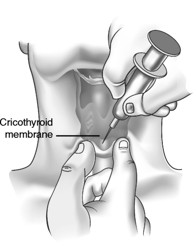

Fig. 67.2
Needle cricothyroidotomy
In children younger than 10–12 years, needle cricothyroidotomy is the preferred method for establishing an emergency airway, since the CT membrane is small and can be difficult to locate quickly. Surgical cricothyroidotomy may easily damage the larynx in this age group, with a higher incidence of postoperative airway complications. In young children, needle cricothyroidotomy should be converted to tracheostomy as soon as feasible.
A large bore needle with catheter (e.g., 14 gauge) attached to a syringe partially filled with water or saline should be immediately accessible in any potential difficult airway situation. Palpation of midline landmarks should allow identification of the space between the thyroid and cricoid cartilages. The needle with attached syringe is advanced through the skin at a 45 ° angle pointing inferiorly. When the needle pushes through the CT membrane, aspiration will reveal air bubbles within the syringe. The needle is withdrawn, leaving the cannula in place. Oxygen tubing is then connected to the cannula, and oxygen can be delivered at a high flow rate of 10–12 l per minute. Use of a Y-connector will allow periodic release of some carbon dioxide.
Surgical Cricothyroidotomy
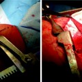
Equipment should be easily accessible and bundled together, avoiding delay. Suggested supplies are listed in Table 67.1. Surgical cricothyroidotomy is performed as follows (Fig. 67.3a–d):
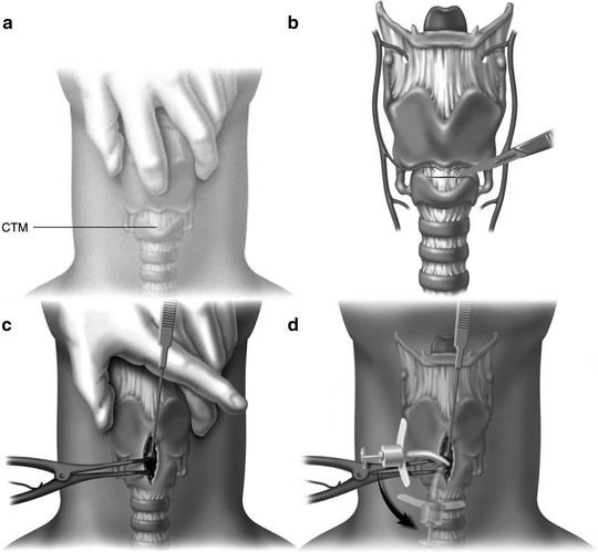
Table 67.1
Equipment for emergency cricothyroidotomy
Scalpel with no. 15 blade |
Hemostats ×2 |
Cricoid hook |
14-g needle with cannula, 6-cc syringe with saline |
Trousseau dilator or Kelley clamp |
Cuffed no.4 tracheostomy tube (previously tested cuff) |
Cuffed no. 5 endotracheal tube (previously tested cuff) |
Gauze |
Betadine, surgical drapes |

Fig. 67.3
Technique for surgical cricothyroidotomy. (a) Neck preparation, positioning, and landmark identification. (b) Skin and cricothyroid membrane incisions. (c) Exposure and dilation of cricothyroid membrane opening. (d) Insertion of an appropriate cannula (From Hagberg CA. Surgical airway. In: Benumof’s Airway Management. Philadelphia: Elsevier; 2007; p. 686–90. Reprinted with permission from Elsevier)
A. Neck Preparation, Positioning, and Landmark Identification
Sterile technique should be observed as much as possible with the application of and appropriate antiseptic solution to the patient’s anterior neck, from the angle of mandible to sternal notch (Fig. 67.3


Stay updated, free articles. Join our Telegram channel

Full access? Get Clinical Tree


