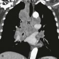
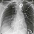
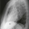
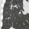
Three relatively recent imaging techniques, computed tomography (CT), ultrasound (US), and magnetic resonance imaging (MRI), have greatly improved thoracic imaging. In all conventional x-ray techniques, the x-ray beam passes through the patient, superimposing all structures in its path onto an x-ray film or detector (projection image, two-dimensional image). Cross-sectional scanning techniques “slice” the patient open, providing a look “inside,” eliminating superimposition. These images are the product of individual digital readings, from multiple angles, synthesized into a digital image. The digital data can be processed to improve tissue contrast and brightness or to view the anatomy in various planes and in three dimensions.


Related posts:


Stay updated, free articles. Join our Telegram channel

Full access? Get Clinical Tree


