Fig. 14.1
Transanal ultrasound showing an internal opening of a transsphincteric fistula, seen as a hypoechoic focus in the intersphincteric space that abuts the internal sphincter, with a small corresponding defect in the internal sphincter
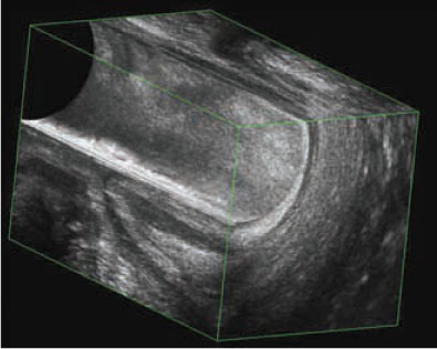
Fig. 14.2
On transanal ultrasound, transsphincteric fistulas are seen as hypoechoic tracts that cross the external sphincter to reach the ischioanal fossa
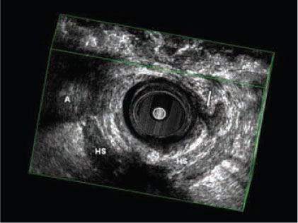
Fig. 14.3
Transanal ultrasound showing an intersphincteric horseshoe extension of a transsphincteric fistula (arrow). A Abscess, HS horseshoe extension
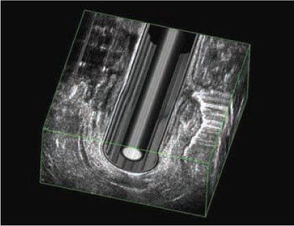
Fig. 14.4
Three-dimensional transrectal ultrasound coupled with the injection of hydrogen peroxide into the external opening of the fistula, showing a complex fistula with extension
To overcome this disadvantage, 3D acquisition, possibly coupled with the injection of hydrogen peroxide into the external opening of the fistula, has become widely adopted (Fig. 14.4) [7]. In 21 patients with a cryptoglandularfistula, West et al. (2003) compared 3D-hydrogen peroxide-enhanced transanal ultrasound with endoanal MRI and surgery. Agreement regarding the classification of the primary fistula tract was 81% for hydrogen-peroxide- enhanced 3D-transanal ultrasound and surgery, 90% for endoanal MRI and surgery, and 90% for hydrogen-peroxide-enhanced 3D transanal ultrasound and endoanal MRI. For secondary tracts, agreement was 67%, 57%, and 71%, respectively, in case of circular tracts and 76%, 81 %, and 71%, respectively, in case of linear tracts. Agreement for the location of an internal opening was 86% for hydrogen-peroxide-enhanced 3D transanal ultrasound and surgery, 86% for endoanal MRI and surgery, and 90% for hydrogen-peroxide- enhanced 3D transanal ultrasound and endoanal MRI (Table 14.1) [8].
Table 14.1
Agreement ratios and k values of 3D hydrogen-peroxide-enhanced ultrasound (HPUS), endoanal magnetic resonance imaging (MRI), and surgery for the classification of perianal fistulas [8]
3D HPUS and surgery | Endoanal MRI and surgery | 3D HPUS and endoanal MRI | |
|---|---|---|---|
Primary tract | 81% | 90% k = 0.47 | 90% |
Circular secondary tract | 67% | 57%
Stay updated, free articles. Join our Telegram channel
Full access? Get Clinical Tree
 Get Clinical Tree app for offline access

|

