Scout view of brain
Lines #1–18 indicate position of sections in the following axial CT series. 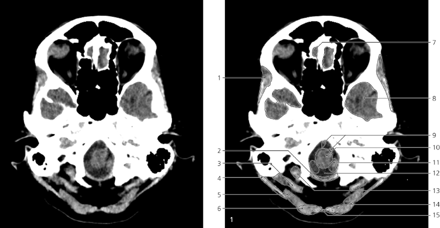
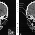
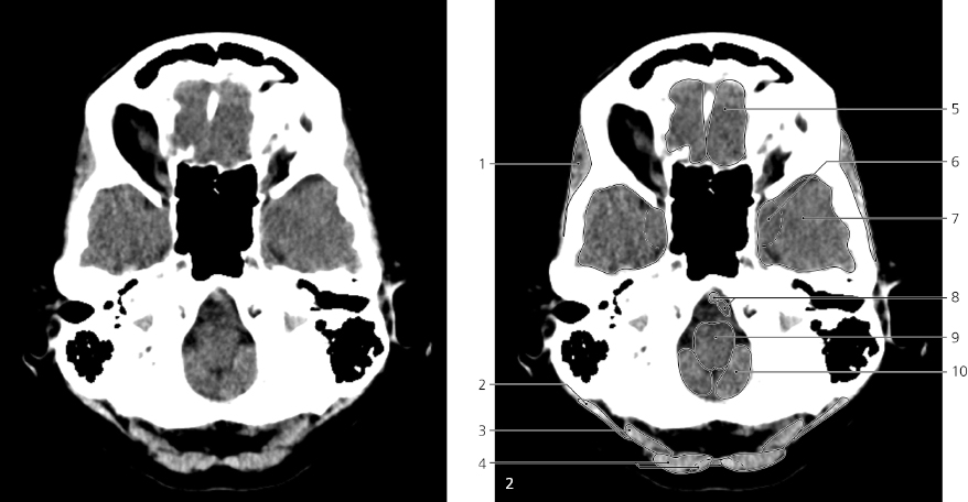
Brain, axial CT
The corresponding image of the skull base is shown on page 207, image #1.
- Temporalis →
- Posterior atlantooccipital membrane
- Rectus capitis posterior major and minor
- Sternocleidomastoideus →
- Splenius capitis and obliuus capitis superior →
- Semispinalis capitis →
- Olfactory bulb
- Temporal lobe →
- Vertebral arteries in cisterna medullaris →
- Medulla oblongata →
- Tonsil of cerebellum →
- Cisterna magna
- Rectus capitis lateralis
- Trapezius →
- Nuchal ligament →
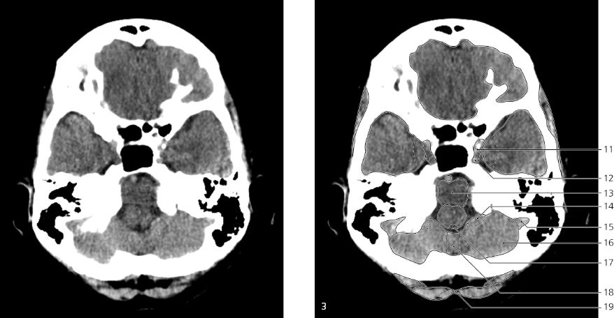
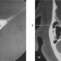
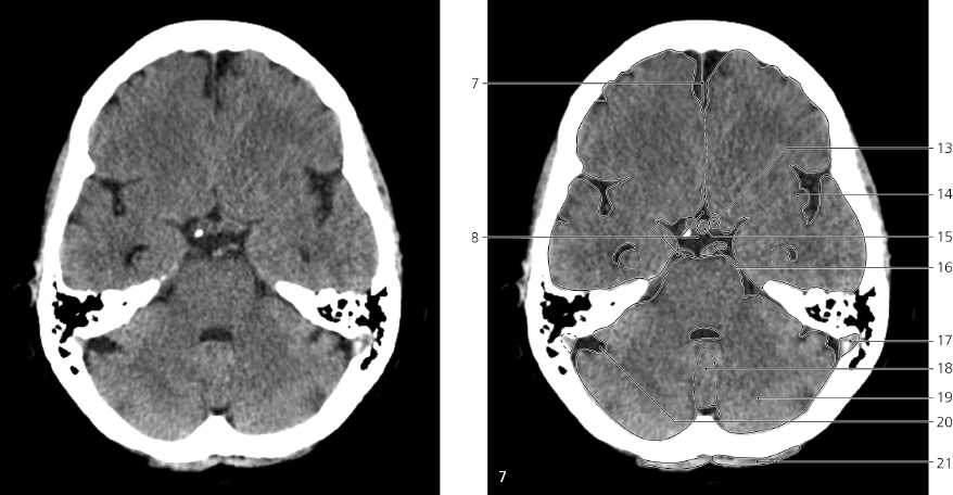
Brain, axial CT
Scout view on page 245. Image #2 of this series is shown in bone settings on page 208. Image #3 is similarly shown on page 209.
- Temporalis ↔
- Sternocleidomastoideus ←
- Splenius capitis and obliquus capitis superior ↔
- Semispinalis capitis and trapezius ↔
- Gyrus rectus
- Uncus of temporal lobe →
- Temporal lobe ↔
- Vertebral arteries in cistern medullaris ←
- Medulla oblongata ←
- Cerebellum (tonsil) ←
- Cavernous sinus →
- Internal carotid artery →
- Pons →
- Flocculus →
- Sigmoid sinus →
- Cerebellar hemisphere →
- Fourth ventricle (obex) →
- Vermis (nodule) →
- Nuchal ligament ←
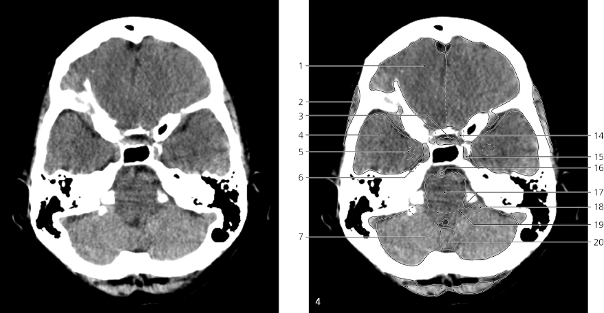
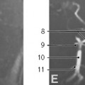
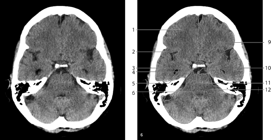
Brain, axial CT
Scout view on page 245. Images #4 and #5 of this series are shown in bone settings on page 210, images #6 and #7.
- Frontal lobe →
- Temporalis ←
- Pituitary gland
- Temporal lobe ↔
- Amygdaloid nucleus
- Trigeminal cave
- Choroid plexus of fourth ventricle
- Falx cerebri ↔
- Infundibulum and pars tuberalis of pituitary gland
- Lateral ventricle (temporal horn) →
- Hippocampus →
- Trigeminal ganglion
- Fourth ventricle ↔
- Internal carotid artery (siphon) ←
- Cavernous sinus ←
- Basilar artery in cicterna pontina ↔
- Pons ↔ and cerebellopontine angle/ cistern
- Flocculus ←
- Inferior cerebellar peduncle
- Vermis (uvula) ↔
- Middle cerebellar peduncle →
- Sigmoid sinus ↔
- Horizontal fissure →
- Semispinalis capitis and trapezius ↔
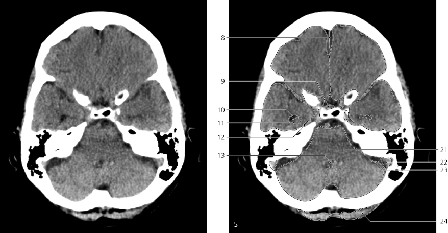
Brain, axial CT
Only gold members can continue reading.
Log In or
Register to continue

Stay updated, free articles. Join our Telegram channel