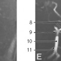
Scout view
- Mandible
- Epiglottis
- Uvula
- Anterior arch of atlas
- Dens axis
- Hyoid bone
- Thyroid cartilage
- Trachea
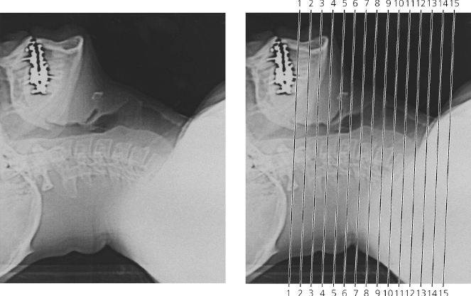
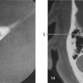
Scout view
Lines #1–15 indicate positions of sections in the following CT-series.Consecutive sections, 10 mm thick 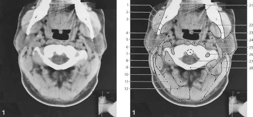
Neck, axial CT
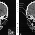
Scout view on previous page
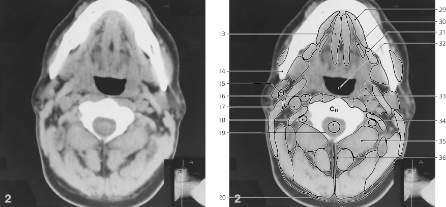
- Masseter
- Medial pterygoid muscle
- Ramus of mandible
- Parotid gland
- Styloid process
- Posterior belly of digastricus
- Sternocleidomastoid
- Obliquus capitis inferior
- Longissimus capitis
- Splenius capitis
- Rectus capitis posterior major
- Semispinalis capitis
- Genioglossus
- Angle of mandible
- Retromandibular vein
- Internal carotid artery
- Internal jugular vein
- Vertebral artery
- Spinal cord
- Trapezius
- Artefacts from dental filling
- Tongue
- Uvula
- Longus colli
- Longus capitis
- Foramen transversarium of atlas
- Dens axis
- Posterior arch of atlas
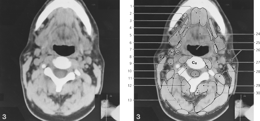
- Mylohyoideus
- Hyoglossus
- Submandibular gland
- Oral part of pharynx
- Lateropharyngeal space
- Levator scapulae, and splenius cervicis
- Obliquus capitis inferior
- Lig. nuchae
Neck, axial CT
Scout view on page 335
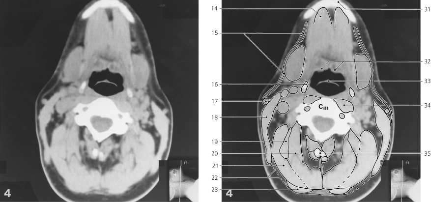
- Geniohyoideus
- Submandibular lymph node
- Mylohyoideus
- Hyoglossus
- Submandibular gland
- Digastricus and stylohyoideus
- External carotid artery (branching)
- Internal carotid artery
- Internal jugular vein
- Vertebral artery
- Intervertebral foramen with spinal nerve
- Spinal cord
- Lig. nuchae
- Digastricus, anterior belly
- Platysma
- Greater cornu of hyoid bone
- External jugular vein
- Sternocleidomastoid
- Longissimus capitis
- Semispinalis capitis
- Splenius capitis
- Superficial lamina of deep cervical fascia
- Trapezius
- Root of tongue
- Oral part of pharynx
- External jugular lymph nodes
- Lateropharyngeal space with vessels, nerves and internal jugular lymph nodes
- Splenius cervicis, and levator scapulae
- Obliquus capitis inferior
- Rectus capitis posterior major
- Mental tuberosity
- Lingual tonsil
- Epiglottis
- Longus colli, and longus capitis
- Spinous process of C II
Only gold members can continue reading.
Log In or
Register to continue

Stay updated, free articles. Join our Telegram channel